Hip Xray Anatomy
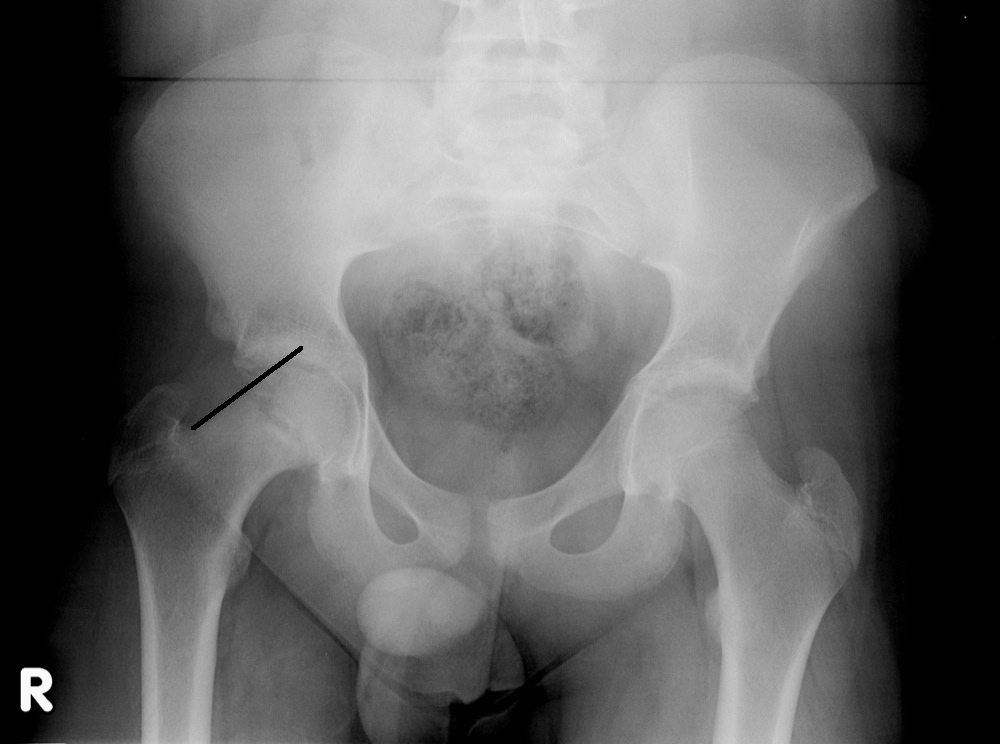
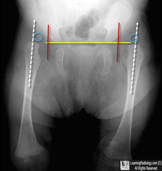
Shentons line is formed by the medial edge of the femoral neck and the inferior edge of the superior pubic ramus. An anteroposterior hip radiograph includes images of both sides of the hip on the same film and projects towards the middle of the line connecting the upper symphysis pubis and anterior superior iliac spine.
 Preoperative Planning Of Total Hip Arthroplasty Intechopen
A standard hip x ray examination generally includes an anteroposterior pa image and a lateral image.

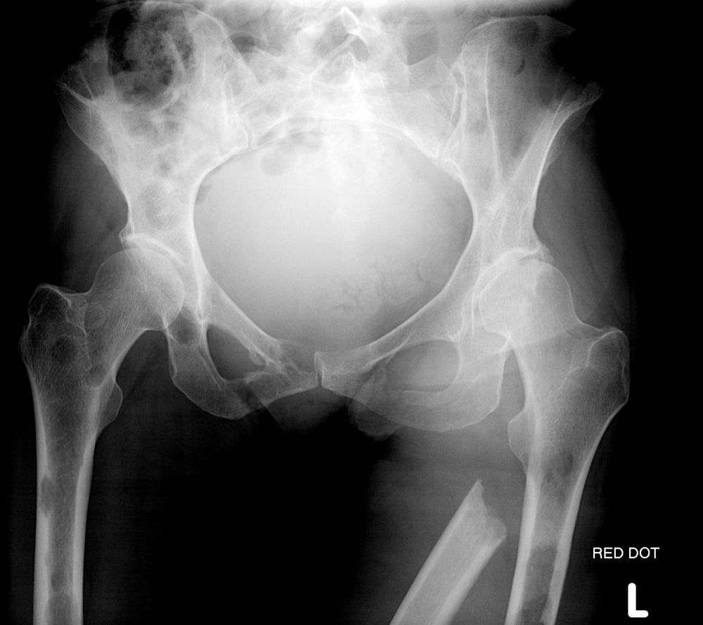
Hip xray anatomy. She is post menopausal and has a borderline osteoporosis of the hips. Notice that the bone in the area of the calcar is much thinner and the cortex of the femoral shaft is much thinner as well. Normal radiographic anatomy of the hip.
In plain radiography x ray anteroposterior and lateral hip radiographs are usually taken. Weight bearing stresses on the hip during walking can be 5 times a persons body weight. The hip joint is one of the largest joints in the body and is a major weight bearing joint.
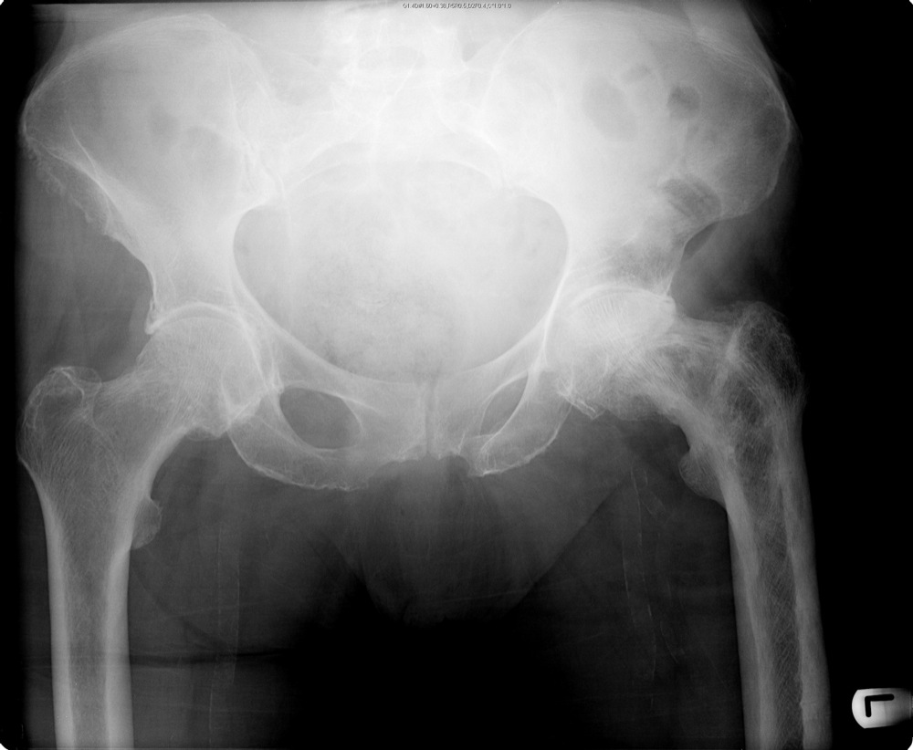
The acetabulum is formed by the three bones of the pelvis the ischium ilium and pubis. The rounded femoral head sits within the cup shaped acetabulum. Ideally the ap image shows both hip joints which strictly speaking makes it a pelvis x ray to allow comparison with the other hip.
The second xray is of the pelvis in a 53 year old female with osteopenia. Normal radiographic anatomy of the hip. Fractures of the femoral neck do not always cause loss of shentons line.
Hip x ray anatomy normal ap. The hip joint is a ball and socket joint that represents the articulation of the bones of the lower limb and the axial skeleton spine and pelvis. Although technically demanding it is the most versatile hip radiograph utilised in trauma bays and general radiography rooms.
A healthy hip can support your weight and allow you to move without pain. Hip horizontal beam lateral view the projection is used to assess the neck of the femur in profile during the investigation of a suspected neck of femur fracture 2. Hip anatomy function and common problems.
Loss of contour of shentons line is a sign of a fractured neck of femur. The distance between the x ray tube and the film should be 12 m. The first xray is of a 35 year old male with no arthritis of the hip.
Normal radiographic anatomy of the hip.
 Film Critique Of The Lower Extremity Part 1
Startradiology
Mr Arthrogram Of The Hip Pathology Pearls And Pitfalls
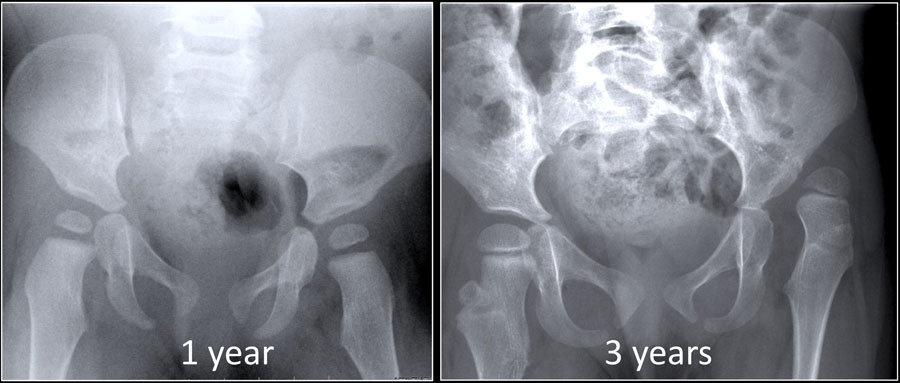
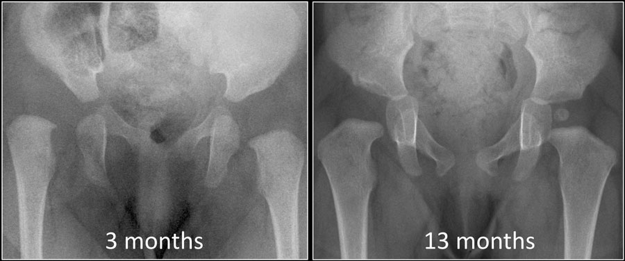 The Radiology Assistant Developmental Dysplasia Of The Hip
 Startradiology
 Hip X Ray Anatomy Normal Ap Shenton S Line Is Formed By The

 Hip Joint Space Measurement In An Anteroposterior Hip

 How To Read Pelvic X Rays International Emergency Medicine
 Anatomy Xray Of The Hip Joint Learn More About Structural
 International Hip Dysplasia Institute

 Film Critique Of The Lower Extremity Part 1
 Ap Hip X Ray Anatomy Diagram Quizlet

Inflammatory Arthritis Of The Hip Orthoinfo Aaos
 Radiographic Anatomy Of Adult Hip Orthopaedicsone Articles
 Radiographic Anatomy Of The Skeleton Hip Anteroposterior

 The Pelvis And Hip
 The Radiology Assistant Developmental Dysplasia Of The Hip
 Normal Radiographic Anatomy Of The Hip Radiology Case
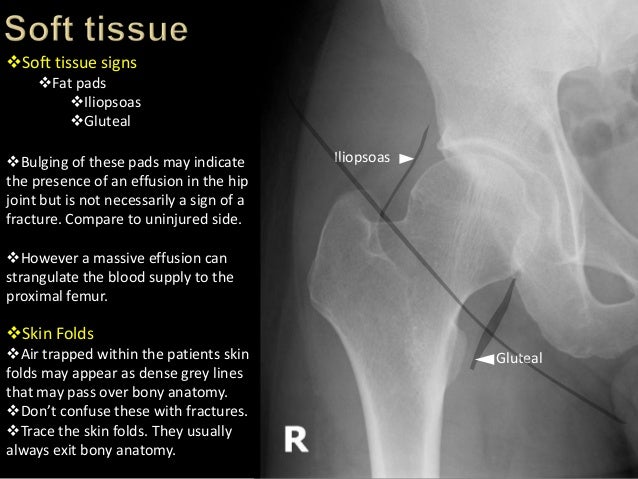 Trauma Image Interpretation Of The Pelvis And Hip

 The Pelvis And Hip
 Radiologic Evaluation Of The Pelvis And Hip Fundamentals
 Dysplasia Xrays
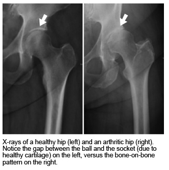 Treating Hip Arthritis Mu Health Care
Hip Resurfacing Orthoinfo Aaos
 Hip Replacement Xray Human Anatomy 3d Illustration Dgi Wire
 Hip Fracture Images Stock Photos Vectors Shutterstock
 The Pelvis And Hip
Belum ada Komentar untuk "Hip Xray Anatomy"
Posting Komentar