Anatomy Of Thumb Tendons
Each finger has 3 phalanges bones and 3 hinged joints. This layer allows the tendons to slide easily through a fibrous tunnel called a sheath.
 Flexor Tendon An Overview Sciencedirect Topics
The extensor apparatus of the thumb is composed of the tendons of the extensor pollicis longus the extensor pollicis brevis the dorsal tendinous hood equivalent of the sagittal band and triangular expansion formed by the oblique fibers given off ulnarly from the tendon of the adductor pollicis and radially from the tendons of the flexor pollicis brevis and abductor pollicis brevis.

Anatomy of thumb tendons. They are located on the posterior aspect of the wrist. Someone with a fractured thumb will usually experience severe pain where the break occurred along with extreme tenderness and a deformed shape of the thumb. The thumb has two of each.
The muscle belly is in the forearm. The extensor digiti minimi straightens the small finger. There are four thumb tendons.
The extensor tendon compartments of the wrist are six tunnels which transmit the long extensor tendons from the forearm into the hand. The flexor digitorum profundus tendon is located under and splits the flexor digitorum superficialis tendon. Basic anatomy of the finger.
One row connects with the ends of the bones in the forearm radius and ulna. Any swelling of the tendons andor thickening of the sheath results in increased friction and pain with certain thumb and wrist movements. Finger movement is controlled by muscles in the forearms that pull on finger tendons.
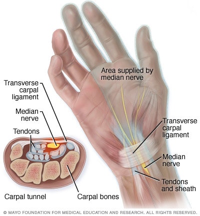
The carpal bones are arranged in 2 interrelated rows. The tendon travels through a tough band or retinaculum at the wrist and then into the hand. Extensor pollicis brevis.
Anatomy of the hand and wrist. Tendons connect muscles to bones. This tendon helps you move the thumb away from the palm to form an open hand.
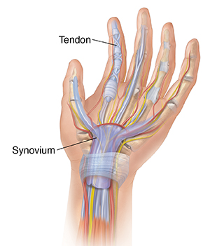
Ligaments connect finger bones and help keep them in place. It attaches to the base of the distal phalanx and flexes the dip. Tendons are covered by a slippery thin soft tissue layer called synovium.
Bones muscles tendons nerves. The wrist links the hand to the arm. 4 figure 1 illustrates the basic anatomy of the finger including joints ligaments and tendons.
The wrist is a complex mechanical system of 8 small bones known as the carpal bones. This tendon travels along the back of the thumb and helps straighten the thumb. It works with the extensor digitorum communis to the small finger.
Swelling numbness coldness and trouble or or inability to move the thumb are other symptoms of a thumb fracturecur near the joints of the thumb. Each tunnel is lined internally by a synovial sheath and separated from one another by fibrous septa. Fingers have a complex anatomy.
Together these combined tendons extend the fingers at the three finger joints. This tendon helps you bend the thumb.
 Anatomy Of Hand Wrist Bones Muscles Tendons Nerves
 Hand And Wrist Anatomy Murdoch Orthopaedic Clinic
 Flexortendonrecovery Learn The Anatomy
 Thumb Hypoplasia Hand Orthobullets
Anatomy 101 Wrist Muscles And Forearm Muscles The
 Structures Of The Hand Tendons Ligaments Teachmeanatomy

 Extensor Tendon Injuries Hand Orthobullets

 Carpal Tunnel Anatomy Mayo Clinic
 Hand And Wrist Anatomical Chart
 Pdf Flexor Tendon Pulleys Thumb Anatomy Zones Langer 2002
 More Insight Into Developing Grip Strength Your Hand Digits
 Hand And Wrist Injuries Part I Nonemergent Evaluation
Applied Anatomy Of The Wrist Thumb And Hand
 Hand And Finger Bones Kirkland Wa Evergreenhealth


Belum ada Komentar untuk "Anatomy Of Thumb Tendons"
Posting Komentar