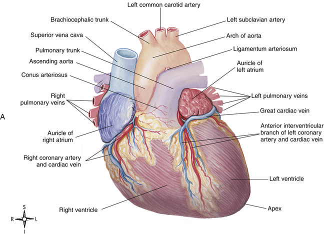
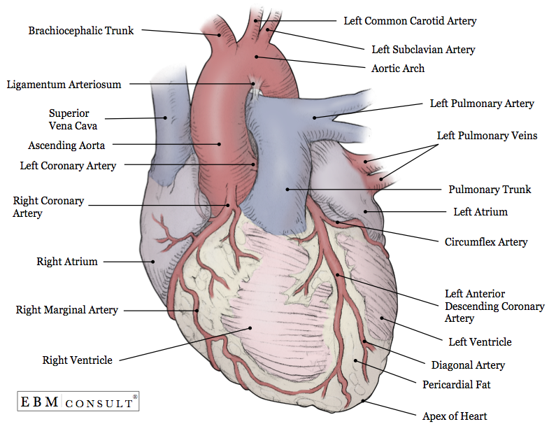
External Anatomy Of Heart
The shape of the heart is similar to a pinecone rather broad at the superior surface and tapering to the apex. Name the artery that carries deoxygenated blood.
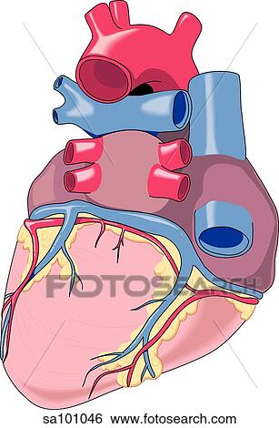
 Heart External Anatomy Drawing Ccp01062 Fotosearch
The human heart is an organ that pumps blood throughout the body through the circulatory system.
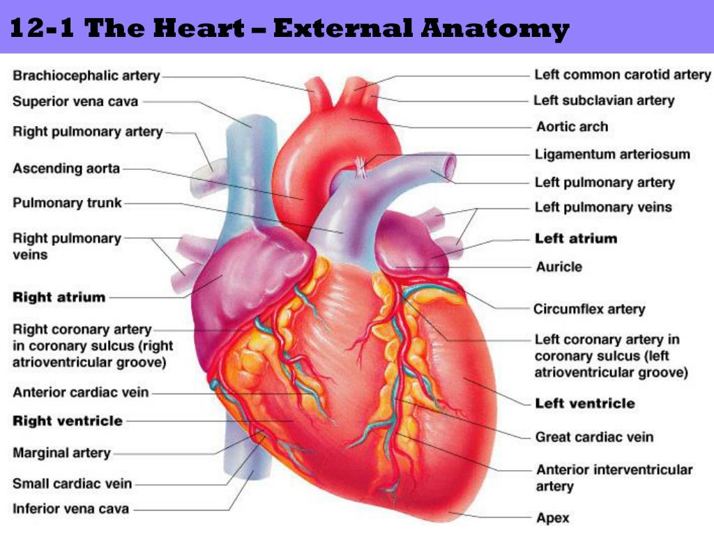
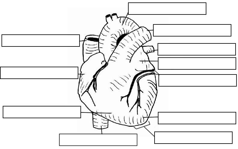
External anatomy of heart. The heart an image of the heart with blank labels attached. The circulatory system lower body image with blank labels attached. The walls and lining of the pericardial cavity are a special membrane known as the pericardium.
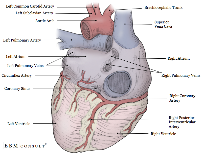
A typical heart is approximately the size of your fist. The right atrium receives blood from the veins and pumps it to the right ventricle. The heart has four chambers.
The lines of reflection between visceral and parietal pericardium form two pericardial sinuses the transverse pericardial sinus and the oblique pericardial sinus. The transverse pericardial sinus lies anterior to the superior vena cava and posterior to the ascending aorta and pulmonary trunk. The heart sits within a fluid filled cavity called the pericardial cavity.
The artery that directs blood from the heart to the lungs carries deoxygenated blood blood that has left its oxygen molecules in many cells so that it is now oxygen poor. External features of heart. Shape and size of the heart.
Images and pdfs. Heart anatomy external the endocardium and subendocardial tissue receive oxygen and nutrients by diffusion or microvasculature directly from the chambers of the heart. The external anatomy of the heart continued d.
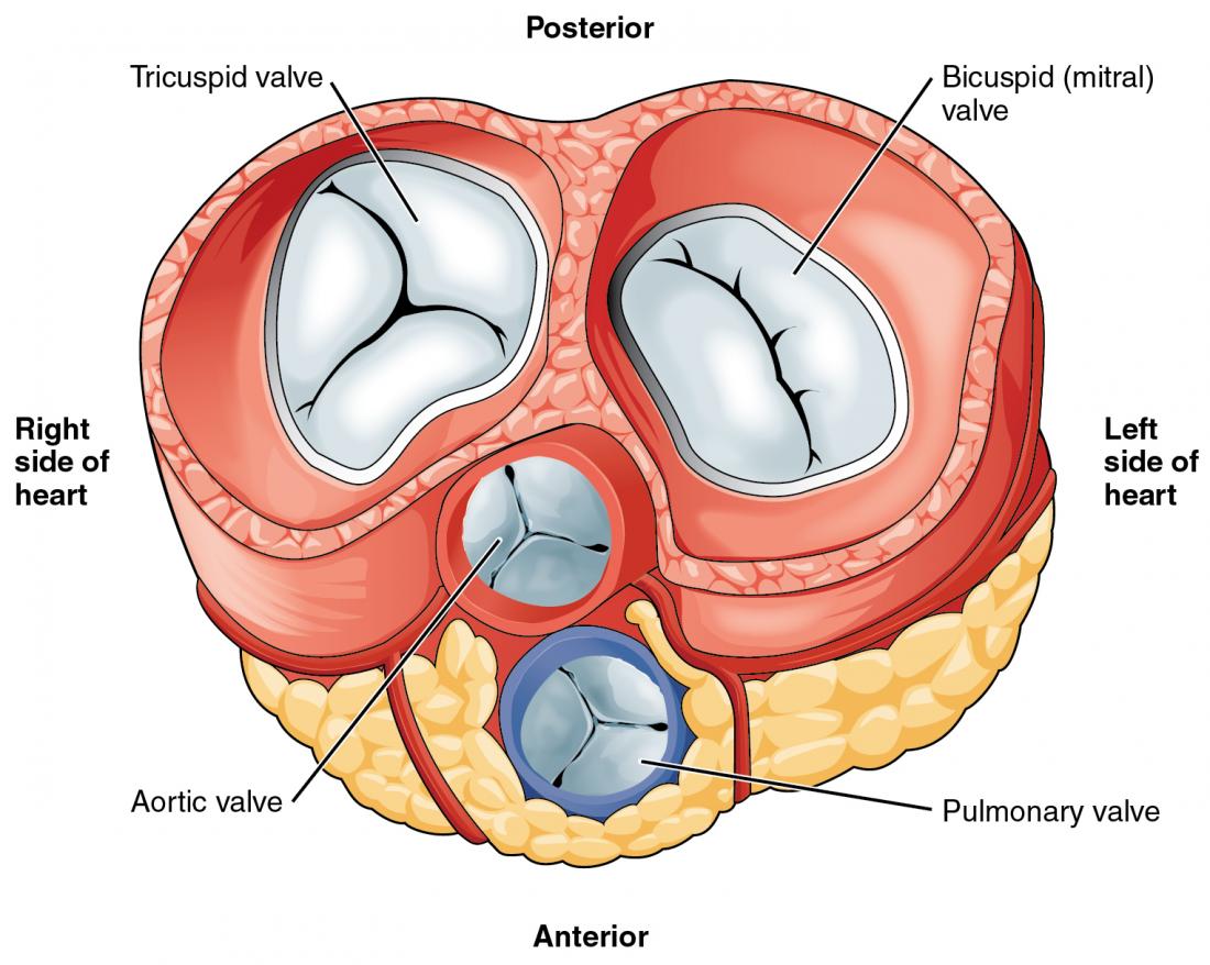
This cross section of the heart shows the right ventricle tricuspid valve left ventricle bicuspid mitral valve left atrium right atrium superior vena cava inferior vena cava aorta aortic valve papillary muscle chordae tendineae and trabeculae carneae. It supplies oxygen and nutrients to the tissues and removes carbon dioxide and other wastes. Pericardium is a type of serous membrane that produces serous fluid to lubricate the heart and prevent friction between the ever beating heart and its surrounding organs.
The circulatory system upper body image with blank labels attached. The remainder is supplied by the coronary vasculature which is primarily embedded in the pericardial fat on the surface of the heart and supplies predominantly the epicardium. 12 cm 5 in in length 8 cm 35 in wide and 6 cm 25 in in thickness.
The right ventricle receives blood from the right atrium and pumps it to the lungs where it is loaded with oxygen. The left atrium receives oxygenated blood from the lungs and pumps it to the left ventricle. The circulatory system a pdf file of the upper and lower body for printing out to use off line.
 Heart Wikipedia
 Heart External Features Anatomy Qa
 Biology Diagrams Images Pictures Of Human Anatomy And
 Anatomical Diagrams Of Heart Heart Failure Online
The Cardiovascular System The Heart
Ppt Cardiovascular Circulatory System Powerpoint
 Posterior View Of The External Anatomy Of The Heart Stock
 External Heart Diagram Reading Industrial Wiring Diagrams
 Anatomy Of The Heart
 The Heart Anatomy Physiology And Function
 Heart And Coronary Arteries Artwork Of The External Anatomy
 Circulatory Systems In Animals Transport Systems In
 Functional Anatomy Of The Cardiovascular System Clinical Gate
 Anatomy Heart External
 Science Source Pig Heart Exterior Anatomy
 Heart 2 External Anatomy Human Anatomy Png Png Free
Search Heart Anatomy Coronary Arteries
 Chapter 19 The Heart Circulatory System Heart Blood
 Anatomy Heart External

 Human Anatomy Physiology Ii
 2 External Features Of The Heart

 Heart External Anatomy Anterior And Posterior Diagram
 Notes Heart And Circulatory System
Belum ada Komentar untuk "External Anatomy Of Heart"
Posting Komentar