Lumbar Spine X Ray Anatomy
Incorrect management of patients with spinal injury may cause or worsen neurological deficit. Lumbar spine 3d reconstruction sacrum and coccyx.
 Presentation1 Pptx Normal Spinal Anatomy
Patient position the radiographs can be performed with the patient in the erect or supine position erect two radiographs.
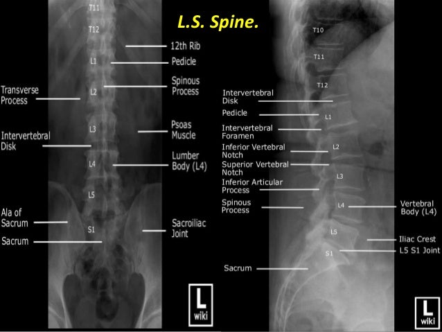
Lumbar spine x ray anatomy. We are pleased to provide you with the picture named lumbar spine and coccyx x ray diagram. Radiological anatomy of the lumbar spine. Ct of the cervical spine.
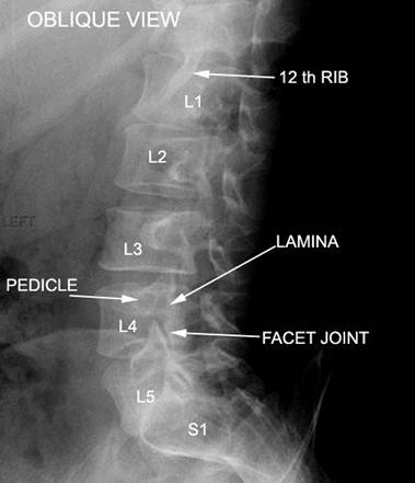
A lumbosacral spine x ray or lumbar spine x ray is an imaging test that helps your doctor view the anatomy of your lower back. Articulations of the facet zygapophyseal joints permit flexionextension and abduction movements. The lumbar spine oblique view is used to visualize the articular facets and pars interarticularis of the lumbar spine.
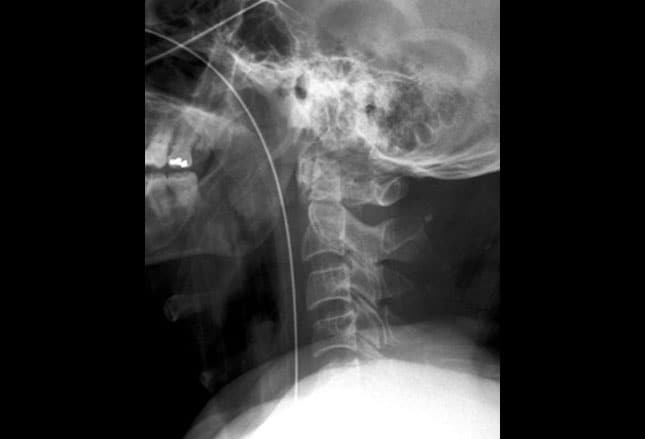
Ct of the craniocervical junction. The lumbar spine consists of five adjacent vertebrae of the lower vertebral column. The plain x ray anatomy and appearances of injuries to both these areas are discussed together.
It uses radiation to make detailed pictures of the bones of your spine. The lumbar spine is made up of five vertebral bones. Therefore patients with suspected spinal injury should be managed by experienced clinicians in accordance with local and national clinical guidelines.
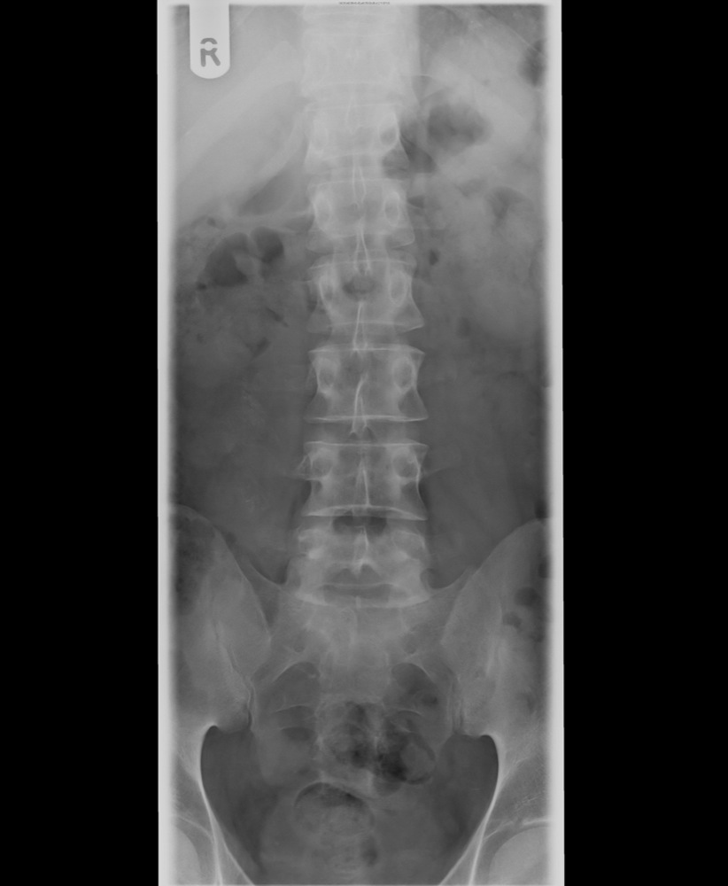
The anteroposterior radiograph anterior aspect shows the vertebral bodies of five lumbar vertebrae their transverse processes spinous and upper and lower joints. Lumbar spine and coccyx x ray diagram in this image you will find disc spaces pedicles facet joints spinous processes inferior articular facest pars interarticularis superior articular facest vertebral body in it. Cervical spine 3d reconstruction mri of the cervical spine.
If your doctor wants to find out whats causing your back or neck pain he may ask you to get a spinal x ray. They participate in the lumbar lordosis a natural curve in the spine that is convex anteriorly.
 Lumbar Spine Operative Neurosurgery
 Lumbar Spine Xray Anatomy Google Search Radiology
 Radiological Anatomy Of The Spine
 Human Xray Of Lumbar Spine Back Anatomy Stock Photo
Cervical Spine Radiographic Anatomy Wikiradiography
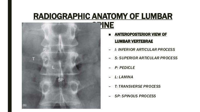
 Radiographic Anatomy Of The Skeleton Lumbar Spine Ap
 Bending Views Of Radiographs Of Lumbar Spine In A 81 Years
 X Ray Mri Lumbosacral Spine A Case Of Low Back Pain Stock
Cervical Spine Radiographic Anatomy Wikiradiography
 How To Read C Spine X Ray International Emergency Medicine
 Ap Lumbar Spine Xray Nutritioneducationcollege Radiology
 Lumbar Spine Anatomy Spine Orthobullets
Cervical Spine Anatomy Diseases And Treatments
 Spine Anatomy And Xray Of Spine Ppt By Dr Pratik
 Thoracolumbar Spine X Rays
Human Xray Of Lumbar Spine Back Anatomy Stock Photo
 Plain Radiographs Of The Lumbar Spine Shows Degenerative
 Technology And Techniques In Radiology X Ray Normal Lumbar
Radiology In Ped Emerg Med Vol 6 Case 13
 X Raying Of The Lumbar Spine Science Publishing Group
 Lateral Thoracic Spine Xray Anatomy Radiology Radiologic
 Interpretations Of The C Spine On Plain Radiography
 The Thoracolumbar Spine
![]() Thoracolumbar Spine X Rays
Thoracolumbar Spine X Rays
 Lumbar Spine X Ray
 Lumbar Spine Oblique Radiology Tutorial
 Lordosis Wikipedia
 Ct Of The Lumbar Spine Anatomy


Belum ada Komentar untuk "Lumbar Spine X Ray Anatomy"
Posting Komentar