Eye Image Anatomy
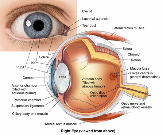
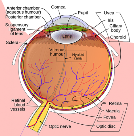
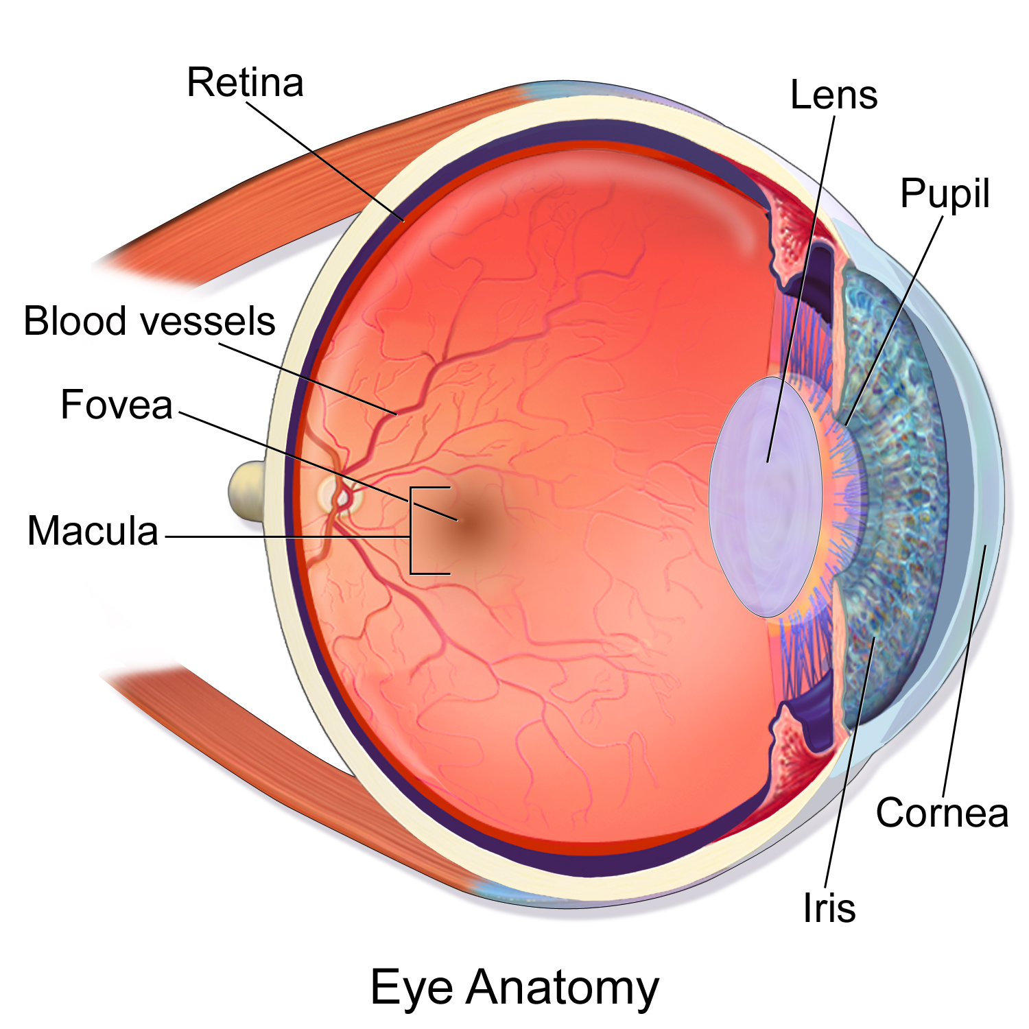
Human eye anatomy seen from above. Anatomy of the eye.
 Eye Anatomy Ocular Anatomy Vision Conditions Problems
Download eye anatomy stock photos.

Eye image anatomy. Oct 8 2017 explore planoeyecares board eye anatomy followed by 387 people on pinterest. Download anatomy stock photos. The cornea is the clear front portion of the eye that processes and focuses light.
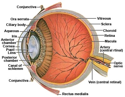
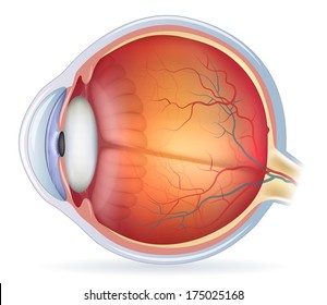
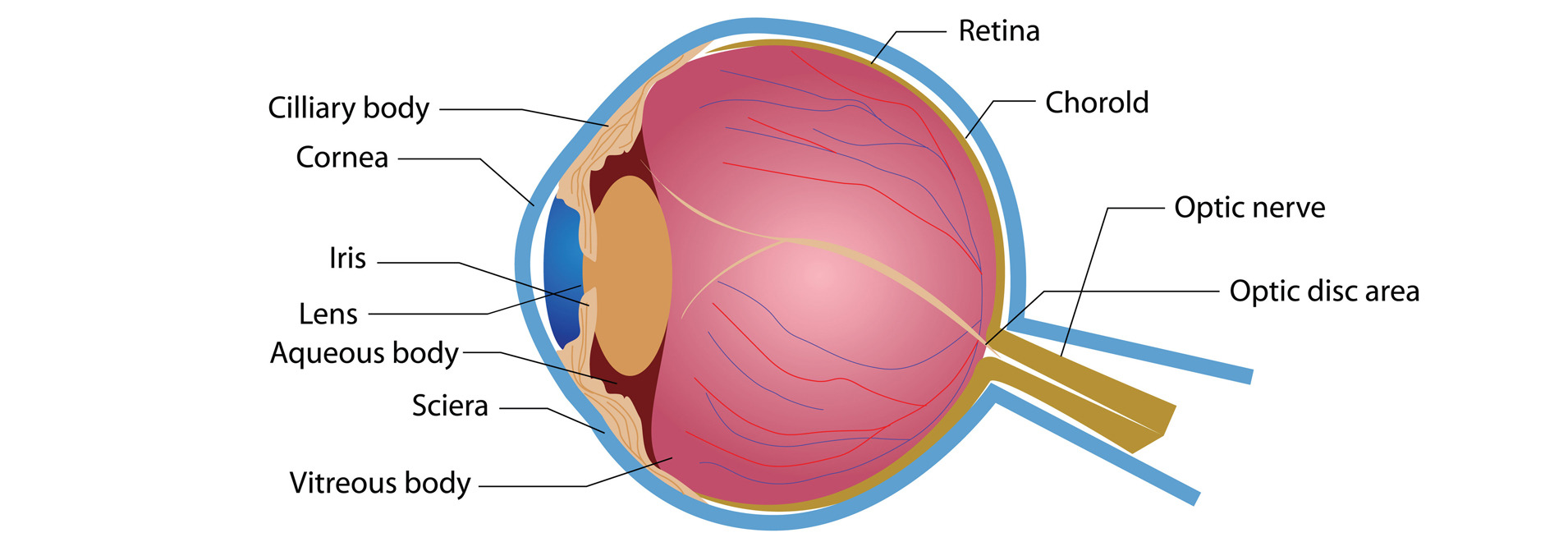
Just as a camera focuses light onto the film to create a picture the eye focuses light onto a specialized layer of cells called the retina to produce an image. The eye has a number of components which include but are not limited to the cornea iris pupil lens retina macula optic nerve choroid and vitreous. Find human eye anatomy stock images in hd and millions of other royalty free stock photos illustrations and vectors in the shutterstock collection.
Just behind the iris and pupil lies the lens which helps focus light on the back of your eye. Light projects through your pupil and. Vision is our window to the outside world.
See pricing plans. The optic nerve then transmits these signals to the visual cortex the part of the brain that controls our sense of sight. Parts of the eye.
26032512 anatomy of the eye cross section normal sight abstract blue. Affordable and search from millions of royalty free images photos and vectors. Clear front window of the eye that transmits and focuses light into the eye.
See more ideas about eye anatomy anatomy and eye facts. Affordable and search from millions of royalty free images photos and vectors. This article explores the anatomy of the eye looking at the different structures of the human eye and their function.
Most of the eye is filled with a clear gel called the vitreous. As we journey through the. The retina acts like an electronic image sensor of a digital camera converting optical images into electronic signals.
The diagrams below show cross sections of the human eyeball. Although the eye is small only about 1 inch in diameter each part plays an important role in allowing people to see the world. Eyes are comprised of many components.
The eye is the organ responsible for vision. Picture of eye anatomy detail the eye is our organ of sight. Picture of eye anatomy detail the eyes are the organs that we use to see.
Thousands of new high quality pictures added every day.
About Basic Eye Anatomy Gem Clinic Glaucoma Eye
Ocular Anatomy And Refractive Error
Eye Anatomy And How The Eye Works
 Royalty Free Human Eye Anatomy Stock Images Photos
Anatomy Of The Eye The Johns Hopkins Wilmer Eye Institute
 Human Eye Ball Anatomy Physiology Diagram
Gross Anatomy Of The Eye By Helga Kolb Webvision
 Eye Anatomy Glaucoma Research Foundation
 Anatomy Of Eye Conditions Pocket Anatomy
 Anatomy Of The Eye Moorfields Eye Hospital
![]() Review Your Eye Anatomy In Order To Understand Eye Disease
Review Your Eye Anatomy In Order To Understand Eye Disease
 Laminated Eye Anatomical Poster Human Eye Anatomy Chart 18 X 27
 Eye Anatomy And Physiology Pupil Iris Retina Cornea Sclera Lens
Eye From Front Anatomy The Eyes Have It
 Anatomy Of A Normal Human Eye Amdf
Gross Anatomy Of The Eye By Helga Kolb Webvision
 Vision And The Eye S Anatomy Healthengine Blog
 Muscle Identification Eye Anatomy Human Anatomy Anatomy
 Anatomy Of The Eye American Association For Pediatric
 Brain And Eye Anatomy Artwork Poster
Anatomy Physiology Pathology Of The Human Eye
 Anatomy Of The Eye Ophthalmology Patient Education Eanw
 Armenian Eyecare Project Anatomy Of The Eye
Major Ocular Structures Laramy K Independent Optical Lab
 Eye Anatomy Central Florida Retina
 Anatomy Of The Eye Kellogg Eye Center Michigan Medicine


Belum ada Komentar untuk "Eye Image Anatomy"
Posting Komentar