Hip Mri Anatomy
This mri hip joint axial cross sectional anatomy tool is absolutely free to use. The hip anatomy on 3t mr and 3d pictures on these 252 3t mri images over 340 anatomical structures are captioned.
![]() Stanford Msk Mri Atlas C 2019
Stanford Msk Mri Atlas C 2019
This radio anatomy atlas is devoted to the articulation and the hip area in mri.
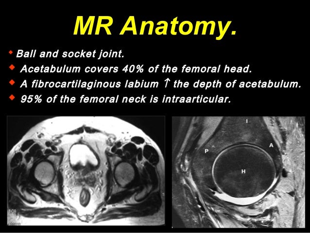
Hip mri anatomy. A magnetic resonance imaging mri is very useful for detecting subtle abnormalities of the hip joint that may not be readily apparent on plain xray. Mri anatomy of the hip. Use the mouse to scroll or the arrows.
Knee shoulder shoulder arthrogram ankle elbow wrist hip contact. The acetabulum is formed by the three bones of the pelvis the ischium ilium and pubis. The hip joint is a ball and socket joint that represents the articulation of the bones of the lower limb and the axial skeleton spine and pelvis.
Click on a link to get t1 axial view t1 coronal view. The rounded femoral head sits within the cup shaped acetabulum. Mri of the hip joint.
This webpage presents the anatomical structures found on hip mri. At the end of the module there are 3d reconstructions of the hip joint hip bone and femur as a recapitulation of musculoskeletal anatomy. Mri of the hip.
Mri of the hip may show normal anatomic variants of the labrum. The variants can be of the labrum itself or associated with the labrum. Use the mouse scroll wheel to move the images up and down alternatively use the tiny arrows on both side of the image to move the images.
The hip joint is a ball and socket type of joint that is also the deepest joint in the body. These anatomic variants can easily be mistaken for pathologic findings on mr images. Stanford bone tumor bayesian network issssr msk lectures for residents ocad msk cases from around the world stanford msk mri atlas has served almost 800000 pages to users in over 100 countries.
Since this joint transfers weight from the upper body to the lower limbs it is subject to a range of problems resulting from faulty weight bearing positions in normal individuals to problems caused by wear and tear in those who are. In the past 10 years mri scans have allowed us to appreciate the subtleties of cartilage and labral degeneration that cause severe hip pain well before obvious osteoarthritis.
 Imaging Anatomy Interactive Pacs Like Atlas Of Radiological
 A Narrative Overview Of The Current Status Of Mri Of The Hip
 Module 2 Lower Extremity Orthopedic Imaging
 Mri Anatomy Of The Hip Review Mri Anatomy Of The Hip
 Mri Female Pelvis Anatomy Axial Im1age 2 Pelvis Anatomy
 Anatomy 24 Pelvic Hip Imaging Flashcards Quizlet
 Mri Of The Thigh Detailed Anatomy
 Hip Joint
 Proximal Iliotibial Band Syndrome Radsource
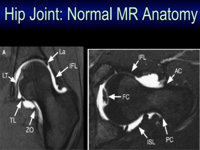
 Mri Anatomy Of The Hip
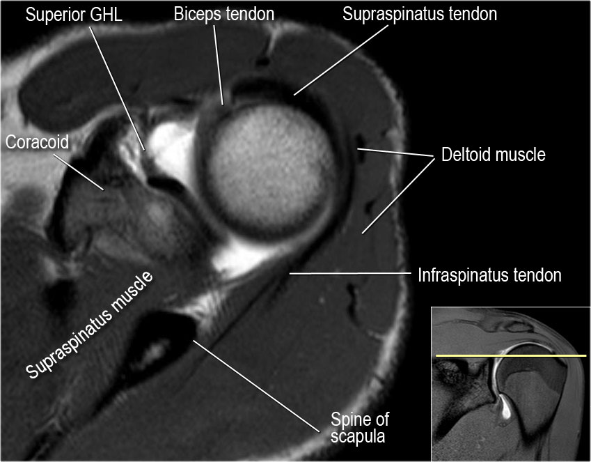
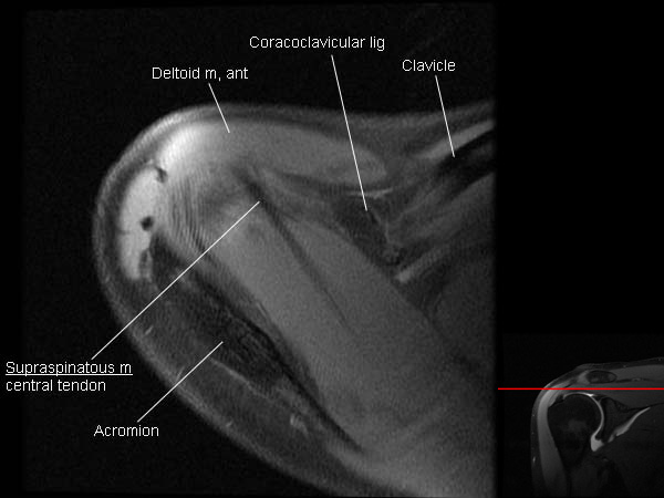
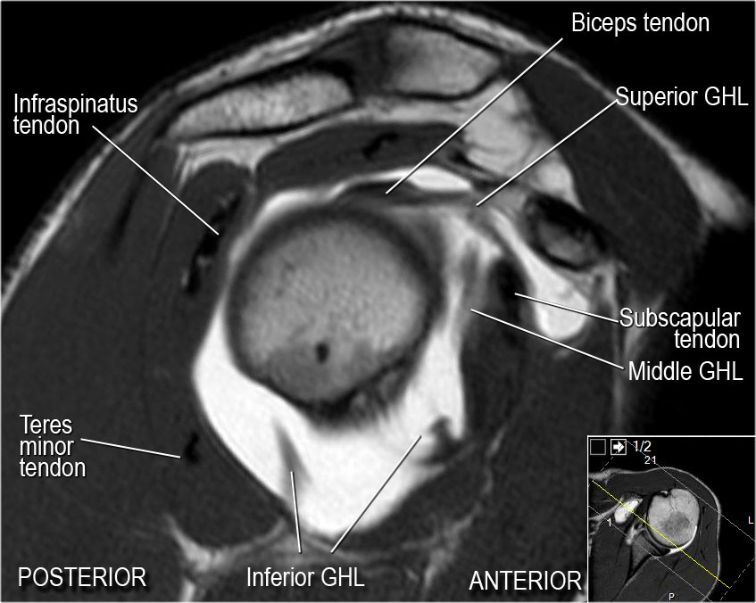 The Radiology Assistant Shoulder Mr Anatomy
 Mri Anatomy Of Hip Joint Free Mri Axial Hip Anatomy
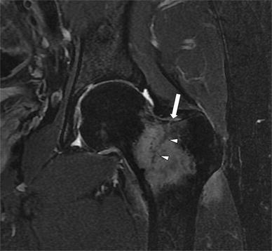 Mri Of The Hip Important Injuries Of The Adult Athlete
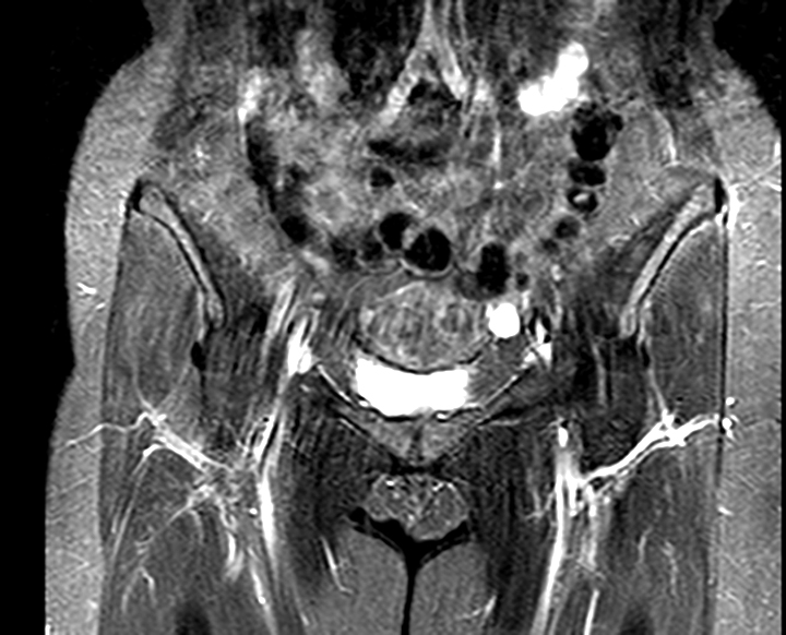
 Anatomy Of Hip Joint Free Mri Coronal Cross Sectional
 Anatomy Of Hip Joint Free Mri Coronal Cross Sectional
 Stanford Msk Mri Atlas 1 0
The Radiology Assistant Shoulder Mr Anatomy
 Hip Joint
 Mobile Mri Of Pelvis Region And Hip Joints A Male 45
 Mri Shoulder Arthrogram Anatomy
 Ultrasonography
 The Hip Anatomy On 3t Mr And 3d Pictures
 Mri Hip Jiont Anatomy Dr Ahmed Eisawy
 Hip Mri Planning Procedure Radtechonduty
 Hip Bursitis Orthoinfo Aaos
 K Anatomy


Belum ada Komentar untuk "Hip Mri Anatomy"
Posting Komentar