Internal Anatomy Of Kidney
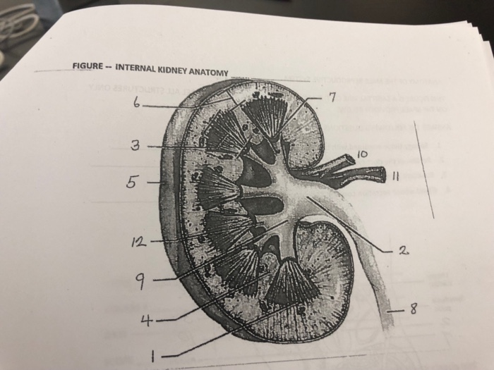
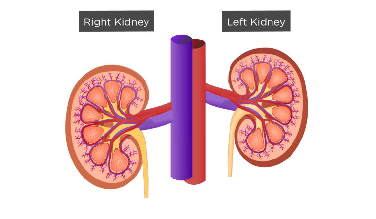
The renal columns are connective tissue extensions that radiate downward from the cortex through the medulla to separate the most characteristic features of the medulla. There are 8 18 renal pyramids in each kidney that on the coronal section look like triangles lined next to each other with their bases directed toward the cortex and apex to the hilum.
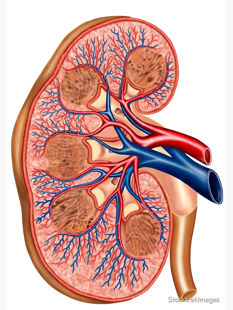
Kidney anatomy encompasses all the internal and external tissue components that collectively form the structure of the kidney.

Internal anatomy of kidney. Renal internal anatomy kidney. Internal anatomy of the kidneys the cortex and medulla make up two of the internal layers of a kidney and are composed of individual filtering units known as nephrons. Learn internal anatomy of the kidney with free interactive flashcards.
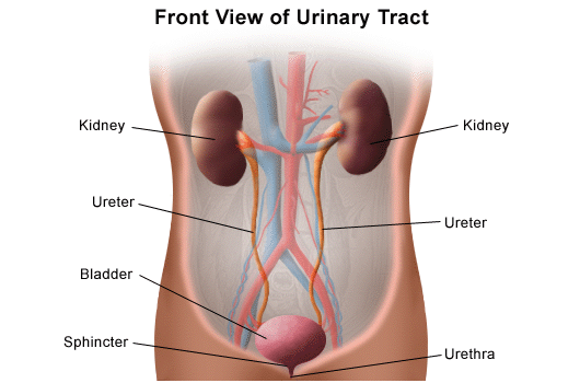
A frontal section through the kidney reveals an outer region called the renal cortex and an inner region called the medulla link. Each kidney is about 4 or 5 inches long roughly the size of a large fist. The kidneys are the main organs of the urinary system and are primarily responsible for removing toxins and other metabolic wastes from the blood.
Numerous tubes and blood vessels located in the cortex make it appear light red and somewhat granular. The kidneys are a pair of bean shaped organs on either side of your spine below your ribs and behind your belly. This dark red area medulla is filled with 8 12 prominent renal pyramids.
A frontal section through the kidney reveals an outer region called the renal cortex and an inner region called the medulla figure 2. In the medulla 5 8 renal pyramid s are separated by connective tissue renal columns. The most external region is referred to as the renal cortex.
The renal columns are connective tissue extensions that radiate downward from the cortex through the medulla to separate the most characteristic features of the medulla. Choose from 500 different sets of internal anatomy of the kidney flashcards on quizlet. Internal anatomy a frontal section through the kidney reveals an outer region called the renal cortex and an inner region called the renal medulla figure 2512.
Internal anatomy of the kidney overview the main unit of the medulla is the renal pyramid. Deep to the cortex is the renal medulla.
 Gross Anatomy Of Urinary System Ppt Video Online Download
What Is The Internal Anatomy Of A Kidney Quora

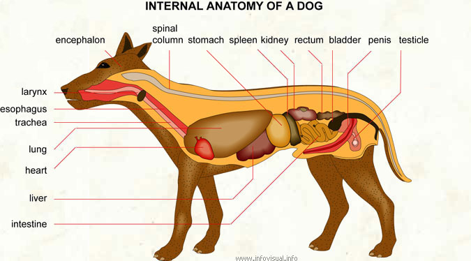 Internal Anatomy Of A Dog Visual Dictionary
![]() Internal Organs Pair Kidneys Isolated Icon Anatomy
Internal Organs Pair Kidneys Isolated Icon Anatomy
 Anatomy Of The Female Urinary Tract Obgyn Key
 Internal Anatomy Of The Kidney Kidney Anatomy Human
:background_color(FFFFFF):format(jpeg)/images/library/10926/renal-arteries_englishAA.jpg) Kidneys Anatomy Function And Internal Structure Kenhub
Kidneys Anatomy Function And Internal Structure Kenhub
 Cross Section Of Internal Anatomy Of Kidney Spiral Notebook
 Pictures Kidneys Male Internal Organs Anatomy 3d
 The Kidneys Boundless Anatomy And Physiology
 Excretory System Internal Structure Of Kidney
 Kidney Stone Disease Poster Art Print Poster
 Human Internal Organs Anatomy Cartoon Vector Stock Image
 Kidney Embroidery Design Science School Nurse Biology Medic Organs Anatomy 169b
 Internal Anatomy Of Kidney Diagram Quizlet
 Internal Anatomy Of The Kidney Purposegames
 Anatomy Of Kidney And Nephron Human Anatomy And Physiology
 Cross Section Internal Anatomy Kidney On Royalty Free
 Kidney And Bladder Urinary System Internal Organs Anatomy Body
 The Internal Anatomy Of The Kidney Anatomy Adipose Tissue
 Kidney Bladder Urinary System Internal Organs Anatomy Body
 Anatomy Of The Human Kidney Cut To Show Internal Structures
Internal Structure Of The Kidney



Belum ada Komentar untuk "Internal Anatomy Of Kidney"
Posting Komentar