Anatomy Of The Cavernous Sinus
Venous blood drains posteroinferiorly to eventually empty into the pytergoid plexuses. The cavernous sinus is made up of very thin walled veins that make up a venous plexus.
 Cavernous Sinus Anatomy
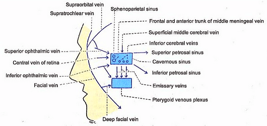
The cavernous sinus receives venous blood from the following.

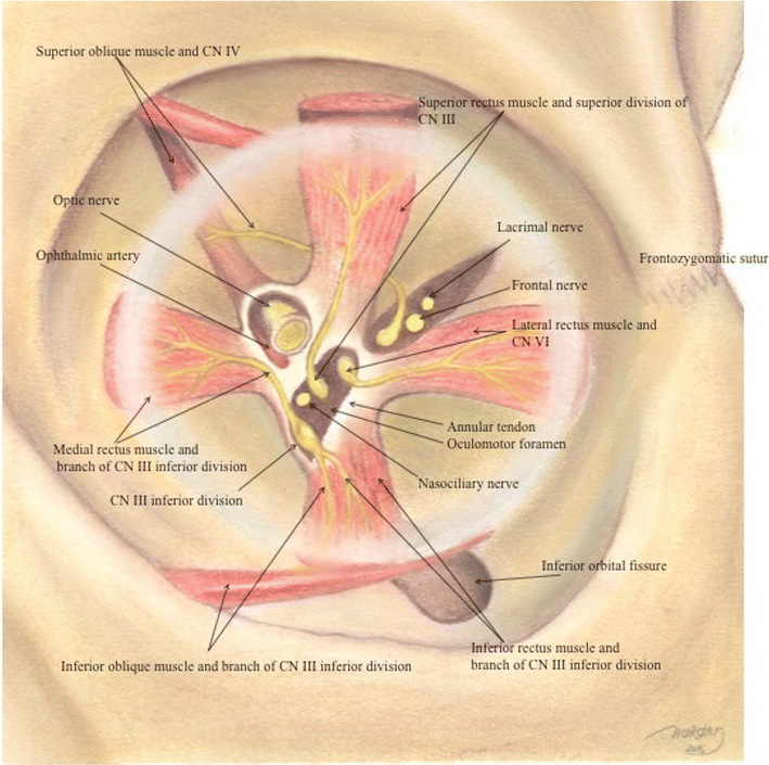
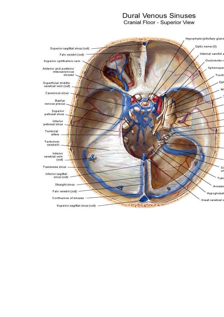
Anatomy of the cavernous sinus. Superior petrosal sinus to the transverse sinus. It is clinically important because of its location its close relationship to several cranial nerves and the internal carotid artery and the complex of veins without valves which drain from and to the paired cavernous sinuses. Anterior superior orbital fissure.
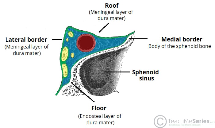
Superior and inferior ophthalmic veins. The cavernous sinus is one of the dural venous sinuses of the head. Medial body of the sphenoid bone.
Posterior petrous part of the temporal bone. Roof meningeal layer of the dura mater. The cavernous sinuses are 1 cm wide cavities that extend a distance.
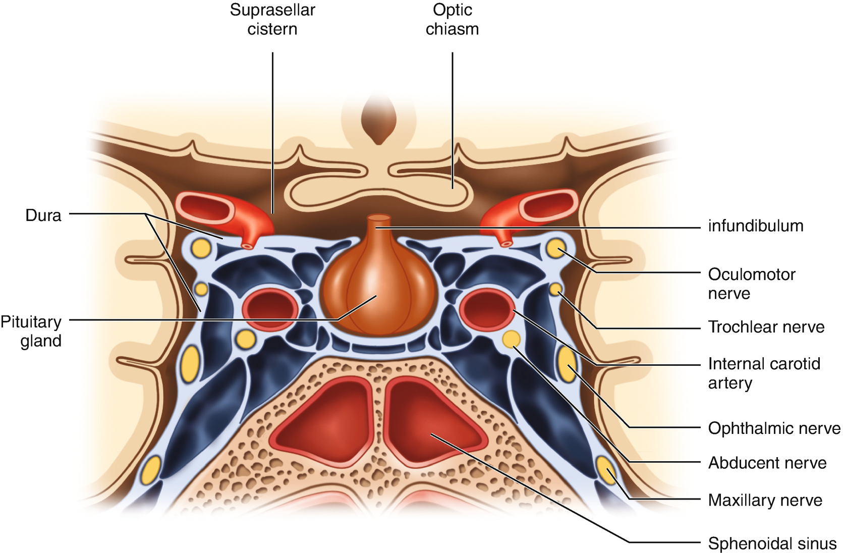
There are numerous structures surrounding the cavernous sinus that are noteworthy. It is a network of veins that sit in a cavity approximately 1 x 2 cm in size in an adult. The cavernous sinus contains the internal carotid artery and several cranial nerves.
2 the carotid siphon of the internal carotid artery and cranial nerves iii iv v branches v 1 and v 2 and vi all pass through this blood filled space. Lateral meningeal layer of the dura mater running from the roof to the floor. The cavernous sinus is a true dural venous sinus and not a venous plexus.
Venous plexus on the internal carotid artery ica to the clival basilar venous plexuses. The borders of the cavernous sinus are as follows. Drainage of the cavernous sinus is via.
Inferior petrosal sinus directly to the jugular bulb. Superior middle cerebral vein. Emissary veins passing through the.
 Operative Management Of Tumors Involving The Cavernous Sinus
 Cavernous Sinus Wikipedia
 Tributaries And Drainage Of Cavernous Sinus Download
 Surgical Anatomy Of The Cavernous Sinus Superior Orbital
 Inferior Ophthalmic Vein An Overview Sciencedirect Topics
 Anatomy Of The Cavernous Sinus Purposegames
Cavernous Sinus Location Drainage Function Human
 Inferior Ophthalmic Vein An Overview Sciencedirect Topics
 Cavernous Sinus Meningiomas Neupsy Key

![]() Cavernous Sinus Anatomy Kenhub
Cavernous Sinus Anatomy Kenhub
 The Cavernous Sinus Contents Borders Thrombosis
 Cavernous Sinus Radiology Reference Article Radiopaedia Org
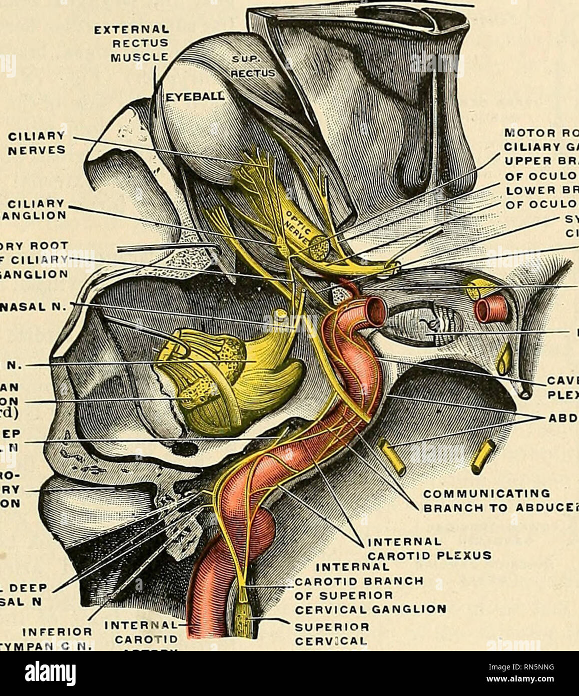 Anatomy Descriptive And Applied Anatomy Cervicocephalic
 Figure 1 From Microsurgical Anatomy Of The Cavernous Sinus
 The Cavernous Sinus Is The Blue Part In Real Life They
 Anatomy Of The Cavernous Sinus And Surrounding Structures
![]() Cavernous Sinus Anatomy Kenhub
Cavernous Sinus Anatomy Kenhub
 Applied Anatomy Of Cavernous Sinus Epomedicine
 Cavernous Sinus An Overview Sciencedirect Topics
 Cavernous Sinus Anatomy Qa
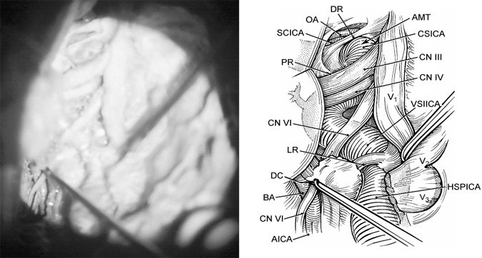 Microsurgical Anatomy Of The Cavernous Sinus
 Cavernous Sinus Syndrome Springerlink
 Mrcp Revision Notes Cavernous Sinus Syndrome
![]() Cavernous Sinus Anatomy Kenhub
Cavernous Sinus Anatomy Kenhub
 Cavernous Sinus Compartments From The Endoscopic Endonasal
 Cavernous Sinus Anatomy
 Cranial Nerves Within The Cavernous Sinus Ami 2018 Meeting
 No More Fear Of The Cavernous Sinuses Sciencedirect
 The Cavernous Sinus Contents Borders Thrombosis

 Pituitary Surgery Neurosurgery Stanford Medicine
Belum ada Komentar untuk "Anatomy Of The Cavernous Sinus"
Posting Komentar