Anatomy Of The Hand And Fingers
Bones muscles tendons nerves. Digits that extend from the palm of the hand the fingers make it possible.
 Muscles Of The Hand Wikipedia
The hand can be considered in four segments.
Anatomy of the hand and fingers. Finger movement is controlled by muscles in the forearms that pull on finger tendons. The thumb has two. The thumb has two proximal and distal.
The digits four fingers and a thumb are made up of small bones called phalanges. There are 3 major types of bones in the hand itself including. The most superficial of all dorsal muscles these can be palpated on the dorsum of the hand.
Fingers have a complex anatomy. Anatomy of the hand. Hand and wrist see also anatomy of the joints bones and joints figure 20.
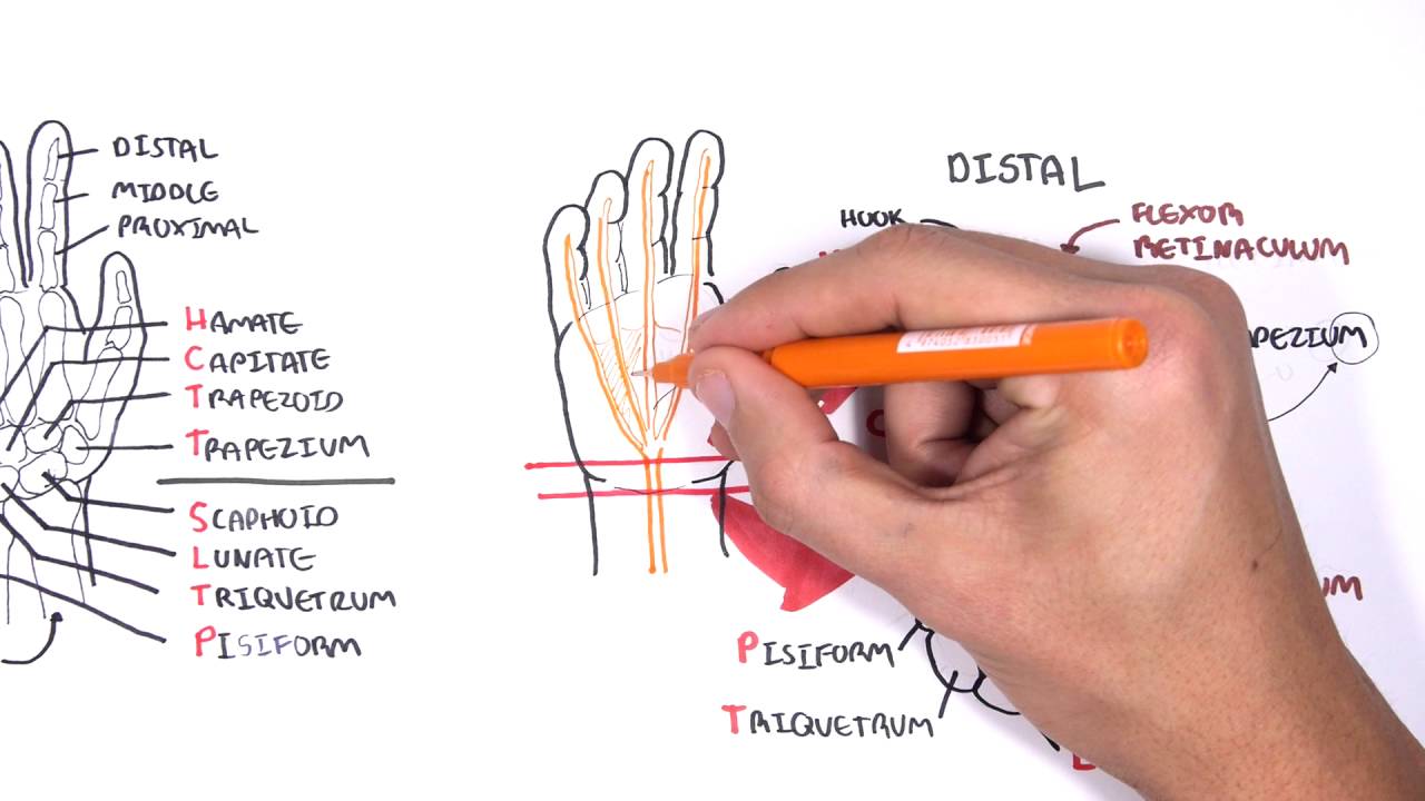
The phalanges singular phalanx the 14 narrow bones that make up the fingers of each hand. Each finger has three phalanges the distal middle and proximal. Anatomy of the hand and wrist.
One row connects with the ends of the bones in the forearm radius and ulna. The carpal bones are arranged in 2 interrelated rows. Choose from 500 different sets of hand and fingers anatomy flashcards on quizlet.
The wrist links the hand to the arm. This is the bottom of the body of the hand. There are four dorsal interossei muscles.
They attach into the extensor hood and proximal phalanx of each finger. The thumb has two of each. Tendons connect muscles to bones.
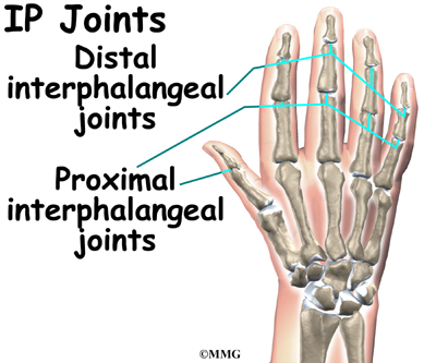
Each interossei originates from the lateral and medial surfaces of the metacarpals. Other bones of the hand are. The next knuckle out toward the fingernail is the proximal inter phalangeal joint pip.
Learn hand and fingers anatomy with free interactive flashcards. Each finger has three phalanges proximal middle and distal. The hand is composed of many different bones muscles and ligaments that allow for a large amount of movement and dexterity.
Each finger has 3 phalanges bones and 3 hinged joints. The back of the hand shows the dorsal venous network a web of veins. The wrist is a complex mechanical system of 8 small bones known as the carpal bones.
This joint commonly is injured in closed fist activities and is commonly known as a boxers fracture. The first and largest knuckle is the junction between the hand and the fingers the metacarpophalangeal joint mcp. Ligaments connect finger bones and help keep them in place.
Abduct the fingers at the mcp joint. The framework of the hand is formed by five metacarpal bones. Picture of finger anatomy.
The 14 bones that are found in the fingers of each hand and also in the toes of each foot. The metacarpals the five bones that comprise the middle part of the hand.
 X Hand Startradiology
 Hand Peace Sign V For Victory Fingers Anatomy Postcard
 Carpal Tunnel Syndrome Cleveland Clinic
 Hand Fractures Treatment Frankfort Wrist Fractures
 Physical Exam Of The Hand Hand Orthobullets
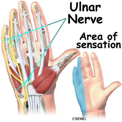
 Hand Anatomy Eorthopod Com
 Hand And Wrist Anatomy Baxter Regional Medical Center
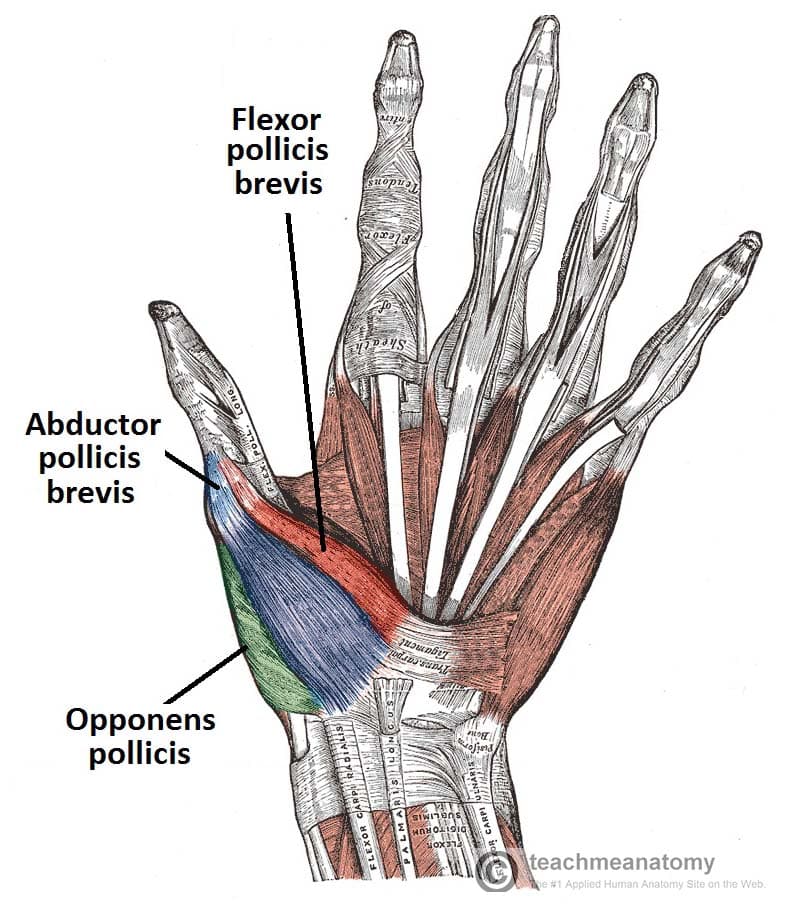 The Muscles Of The Hand Thenar Hypothenar Teachmeanatomy
 Hand Anatomy Orthogate
 Structures Of The Hand Tendons Ligaments Teachmeanatomy
 Intrinsic Hand Muscles Msk Medbullets Step 1
 The Hand Muscles Episode 24 Anatomy On The Go
 Hand Wikipedia
 Hand Anatomy Midwest Bone Joint Institute Elgin Illinois
 Hand And Finger Bones Kirkland Wa Evergreenhealth
Hand Anatomy Eorthopod Com
 Wrist Hand Atlas Of Anatomy
 Your Finger Joint Pain Is Probably Caused By Arthritis
 Hand Anatomy Healthengine Blog
Anatomy Of The Hand Johns Hopkins Medicine
Hand Anatomy Tuned In Guitar Lessons
 A Sixth Finger Can Prove Extra Handy Science News For Students
Understanding Infectious Tenosynovitis Of The Finger Hand
 Hand Anatomy Overview Bones Blood Supply Muscles Geeky
 Anatomy Of The Hand Team Bone
 Clinical Anatomy Hand Wrist Palmar Aspect Flexors
 Better Information Better Health Hand Anatomy Muscle

Belum ada Komentar untuk "Anatomy Of The Hand And Fingers"
Posting Komentar