Anatomy Of Patella
The patella bone is more commonly referred to as the kneecap. The patella is commonly referred to as the kneecap.
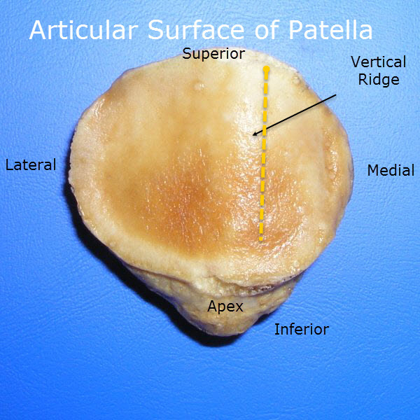 Anatomy Of Patella Bone And Spine
The patella bone is a sesamoid bone that is a part of the appendicular skeleton.
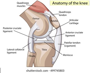
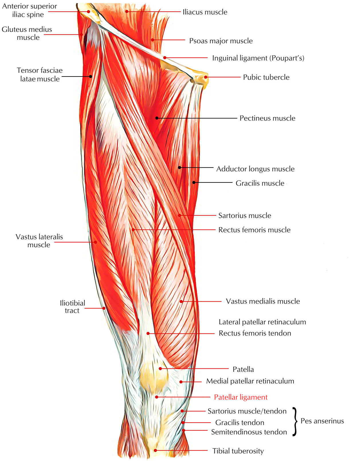
Anatomy of patella. The apex of the patella is situated inferiorly and is connected to the tibial tuberosity by the patella ligament. Determination of side of patella a triangular with the apex of the triangle directed downwards. The bone originates from multiple ossification centres that develop from the ages of three to six which rapidly coalesce.
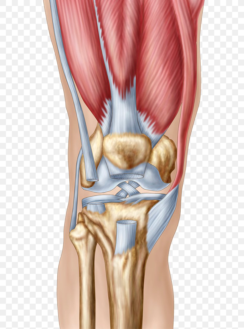
The patella is held in place by muscles the lower end of which surrounds the patella and is then attached to the upper part of the tibia shin by patellar tendons. The upper three fourths of the posterior surface is smooth and articular. Patella anatomy patella bone anatomy video and notes.
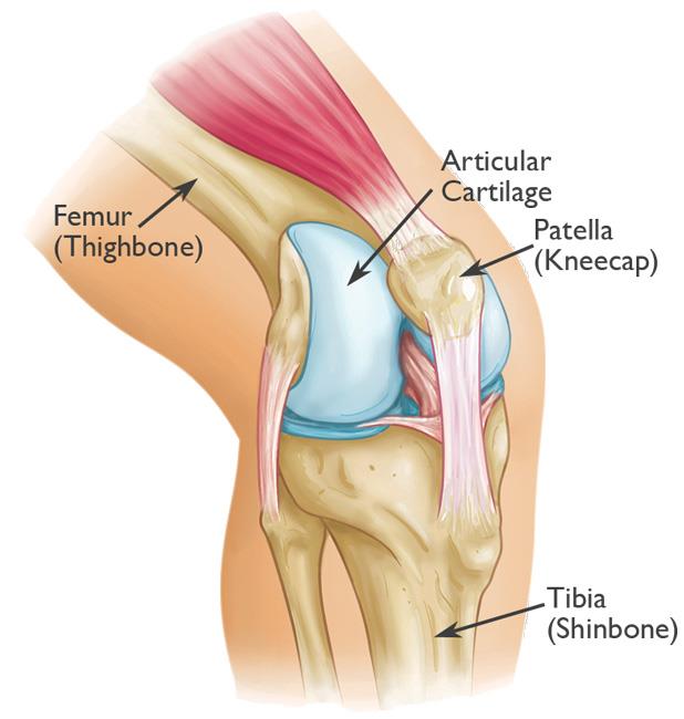
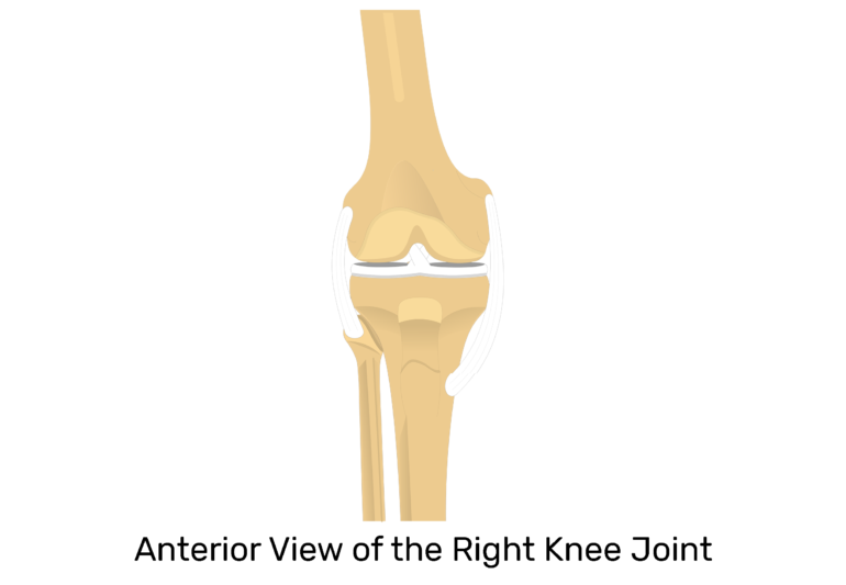
The patella is the technical name for the kneecap the triangular shaped bone at the front of the knee joint. The patella is the largest sesamoid bone in the body and it lies within the quadriceps tendon in front of the knee joint. It is a small freestanding bone that rests between the femur thighbone and tibia shinbone.
The femur has a dedicated groove along which the kneecap slides. The patella protects the knee joint. The base forms the superior aspect of the bone and provides the attachment area for the quadriceps tendon.
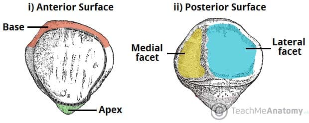
As a form of protection both bones also contain cartilage strong flexible tissue in the areas near the patella. The anterior surface is rough and nonarticular. The patella is a thick flat triangular bone with its apex pointing downwards.
The patella has a triangular shape with anterior and posterior surfaces.
 Patella Bones Location Function
:watermark(/images/logo_url.png,-10,-10,0):format(jpeg)/images/anatomy_term/patella-5/dDgJWgQNyzceGp9ye8CfmA_Patella_01.png) Patella Anatomy Function And Clinical Aspects Kenhub
Patella Anatomy Function And Clinical Aspects Kenhub
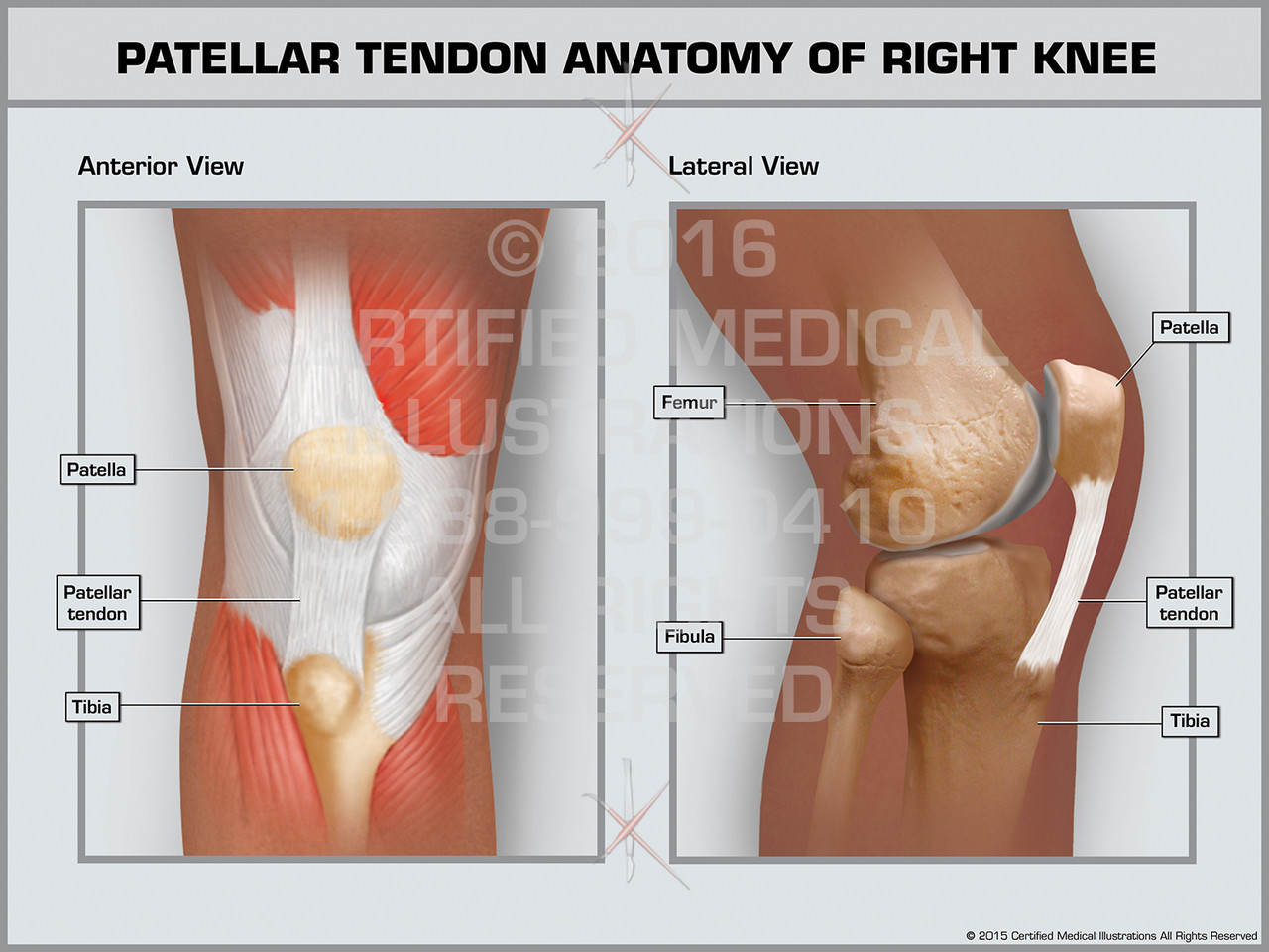 Patellar Tendon Anatomy Of Right Knee Print Quality Instant Download
Patellofemoral Arthritis Orthoinfo Aaos
 Amazon Com Emvency Mouse Pads Pain Patella Knee Joint
:max_bytes(150000):strip_icc()/Blausen_0597_KneeAnatomy_Side-5bbfb48fc9e77c0051f630c3.jpg) What Is The Patellofemoral Joint
What Is The Patellofemoral Joint
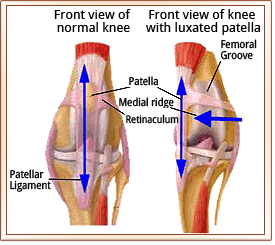
-Luxation-Anatomy-Chart.jpg) Patella Medial Luxation In Dogs Vetlexicon Canis From
Patella Medial Luxation In Dogs Vetlexicon Canis From
 The Patella Surface Anatomy Functions Dislocation
 Patella An Overview Sciencedirect Topics
 Patella Images Stock Photos Vectors Shutterstock
 Anatomy Of The Peripatellar Fat Pads The Suprapatellar Fat
 Knee Joint Anatomy Pictures And Information
 Patellar Fractures Broken Kneecap Orthoinfo Aaos
 Patella Tendon Rupture Core Em
/188058334-crop-56aae7425f9b58b7d0091480.jpg) What Is Causing Your Knee Pain
What Is Causing Your Knee Pain
 Easy Notes On Patellar Ligament Learn In Just 4 Minutes
 Patellar Luxation Metropolitan Veterinary Associates
 Anatomy Of The Knee Baxter Regional Medical Center
 Stuck In The Middle Patellar Tracking And Pain
 Anatomy Of Patella Bone And Spine
 Knee Human Anatomy Function Parts Conditions Treatments
 Knee Pain Human Anatomy Patella Png 606x1105px Watercolor
Soft Tissue Knee Patient Information Gavin Mchugh
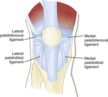 Patellofemoral Joint Physiopedia
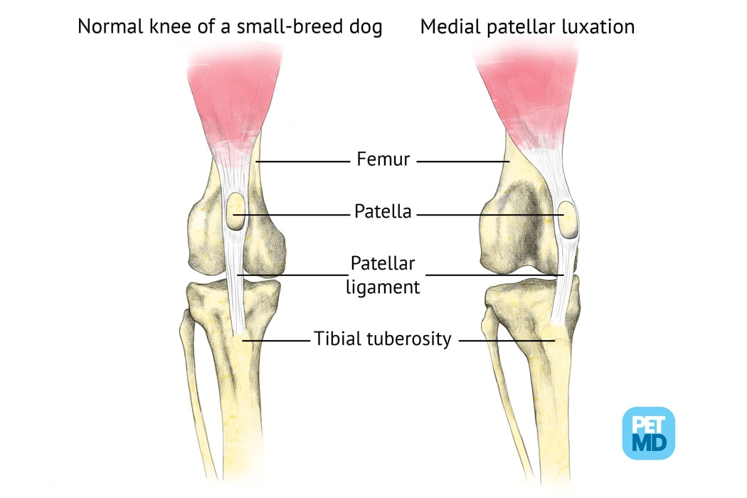 Patellar Luxation In Dogs Medical Diagram
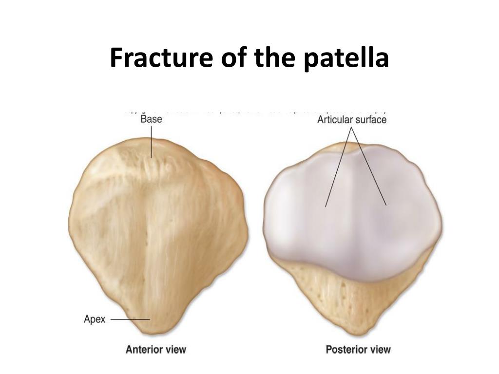 Ppt Fracture Of The Patella Powerpoint Presentation Free
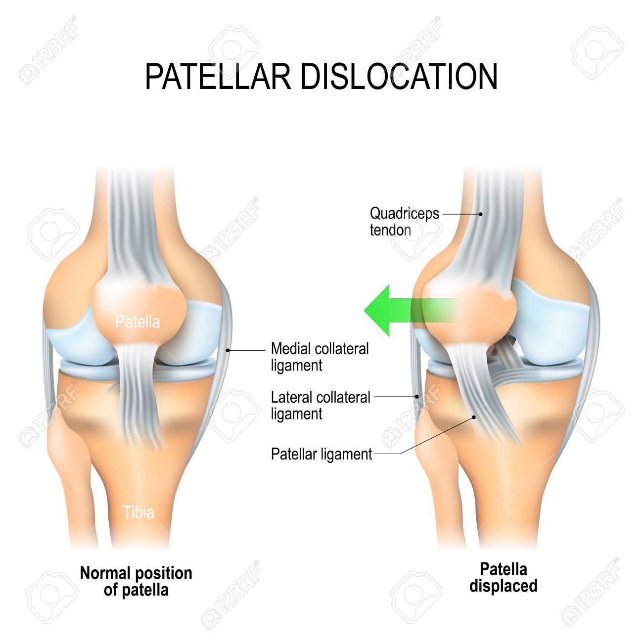 Patellar Dislocation Normal Position Of Kneecap And Patella
 Patella Bone Anterior And Posterior Views
Patellar Femoral Pain Patellar Tendinitis Renew Physical
 Welcome To Madison Street Animal Hospital Patella Surgery


Belum ada Komentar untuk "Anatomy Of Patella"
Posting Komentar