Brain Anatomy Labeled
Structure descriptions were written by levi gadye and alexis wnuk and jane roskams. The nervous system the anatomy of the nervous system.
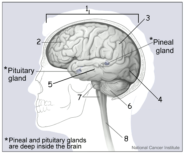 Nervous System Label The Brain
The nervous system an image of the nerves of the lower body with blank labels attached.
Brain anatomy labeled. The nervous system an image of the nerves of the upper body with blank labels attached. Non contrast sagittal ct head. Use the mouse scroll wheel to move the images up and down alternatively use the tiny arrows on both side of the image to move the images.
The brain is composed of the complex network of billions of neurons that are arranged in a specific pattern which is vital to the essential functioning of this organ. In the above picture. Identify the region labeled 2.
Angiogram axial ct head. Practice lab practical on human brain anatomy. This mri brain cross sectional anatomy tool is absolutely free to use.
Identify the space labeled 4. The basal ganglia are a cluster of structures in the center of the brain. 12 cerebrum cerebellum brain stem spinal cord this part of the nervous system moves messages between the brain and the body.
This interactive brain model is powered by the wellcome trust and developed by matt wimsatt and jack simpson. Identify the region labeled 1. Non contrast axial ct head.
The frontal. Non contrast coronal ct head. 11 this brain part controls involuntary actions such as breathing heartbeats and digestion.
Angiogram coronal ct head. Reviewed by john morrison patrick hof and edward lein. The nervous system the physiology of the nervous system.
The part of the brain stem that joins the hemispheres of the cerebellum and connects the cerebrum with the cerebellum. The cerebellum is at the base and the back of the brain. Picture of the brain the cortex is the outermost layer of brain cells.
Anatomy of the brain overview the brain is an amazing three pound organ that controls all functions of the body interprets information from the outside world and embodies the essence of the mind and soul. Regulates sleep and dreams. What do regions 1 2 and 3 comprise.
Identify the space labeled 5. Identify the region labeled 3. The brain stem is between the spinal cord and the rest of the brain.
A thick bundle of nerve fibers that runs from the base of the brain to the hip area running through the spine. Working on the principle of division of labour different parts of brain are specialized for only specific tasks. This article lists a series of labeled imaging anatomy cases by system and modality.
:background_color(FFFFFF):format(jpeg)/images/library/11646/basic-anatomy-of-the-brain_english_copy.jpeg) Parts Of The Brain Learn With Diagrams And Quizzes Kenhub
Parts Of The Brain Learn With Diagrams And Quizzes Kenhub
 Brain Png Transparent Image Labeled Brain Anatomy
Post It Anatomy Of The Brain Anatomical Chart 11 X 17
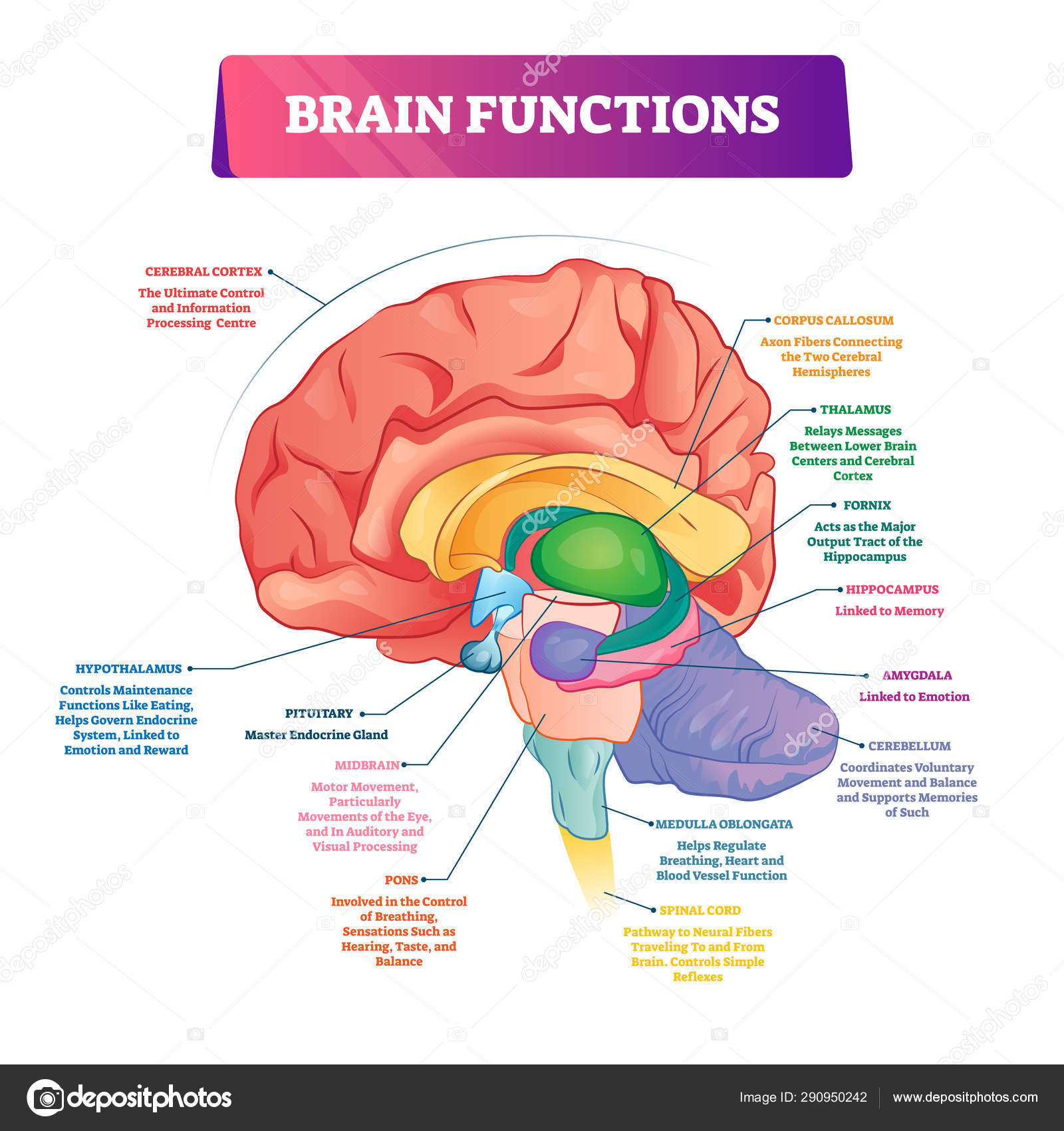
 Brain Functions Vector Illustration Labeled Explanation
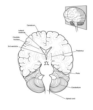 Brain Anatomy Overview Gross Anatomy Cerebrum Gross
 Science Source Brain Anatomy Illustration
 Human Brain Anatomy Regions Labeled Educational Chart Framed Poster 20x14 Inch
 Anatomy Brain Model Brain Anatomy Model Labeled
![]() Colored And Labeled Human Brain Diagram Flat Vector Illustration
Colored And Labeled Human Brain Diagram Flat Vector Illustration
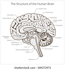 Labeled Brain Anatomy Images Stock Photos Vectors
 Brain Anatomy Labeled A H Diagram Quizlet
 Brain Diagram To Label Wiring Diagram Images Gallery
:background_color(FFFFFF):format(jpeg)/images/library/6721/lateral-views-of-the-brain_english.jpg) Lateral View Of The Brain Anatomy And Functions Kenhub
Lateral View Of The Brain Anatomy And Functions Kenhub
 Anatomical Diagrams Of The Brain
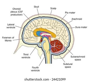 Labeled Brain Anatomy Images Stock Photos Vectors
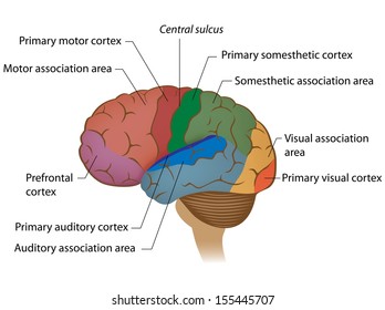 Labeled Brain Anatomy Images Stock Photos Vectors
 Labeled Pictures Of The Brain Labeled Pictures Of The

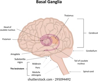 Labeled Brain Anatomy Images Stock Photos Vectors
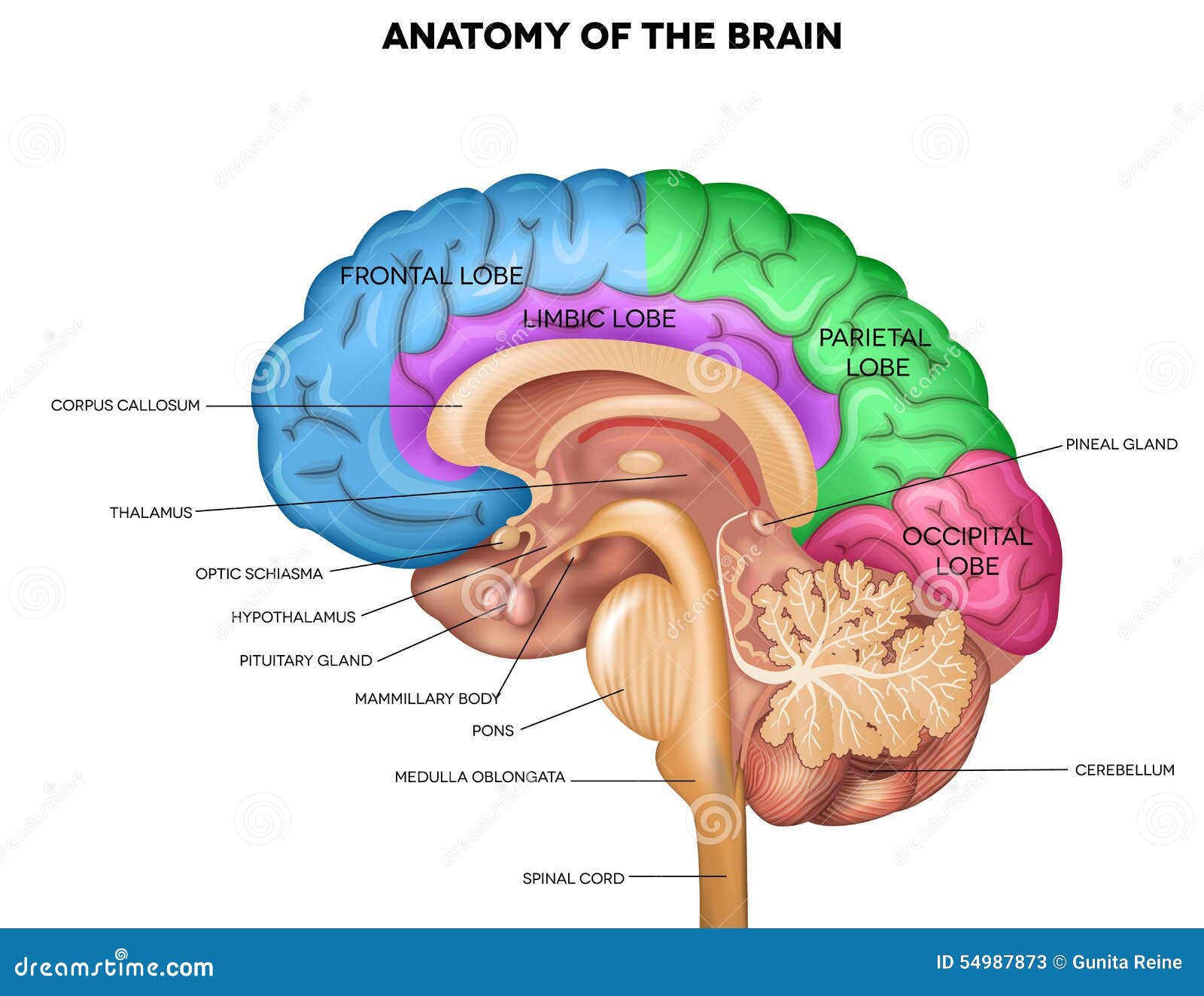 Human Brain Anatomy Stock Vector Illustration Of Normal
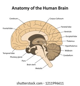 Labeled Brain Anatomy Images Stock Photos Vectors
 Median Section Of Human Brain Diagram Clipart K49070621
 Brain Anatomy Diagrams Brain Anatomy Brain Diagram
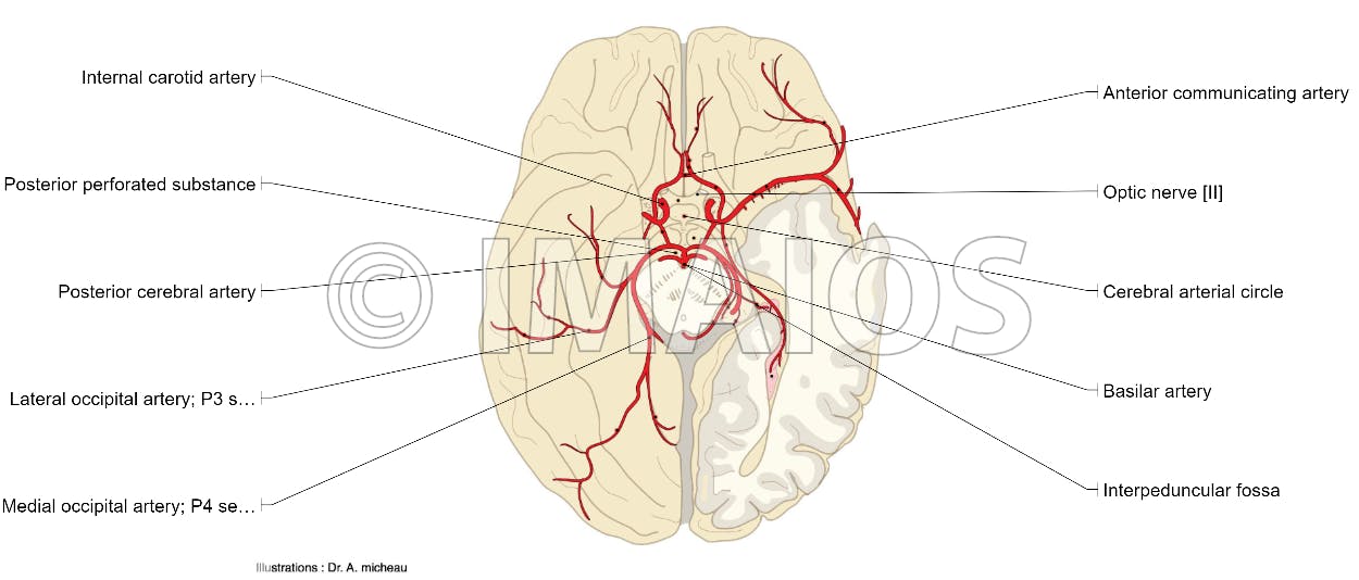 Anatomical Diagrams Of The Brain
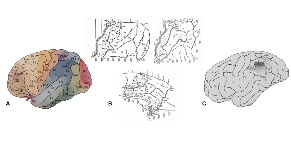 Labeled Brain Anatomy Images National Chimpanzee Brain
 Brain Labeling 2 And Ventricles Anatomy And Physiology
Free Brain Diagram Download Free Clip Art Free Clip Art On


Belum ada Komentar untuk "Brain Anatomy Labeled"
Posting Komentar