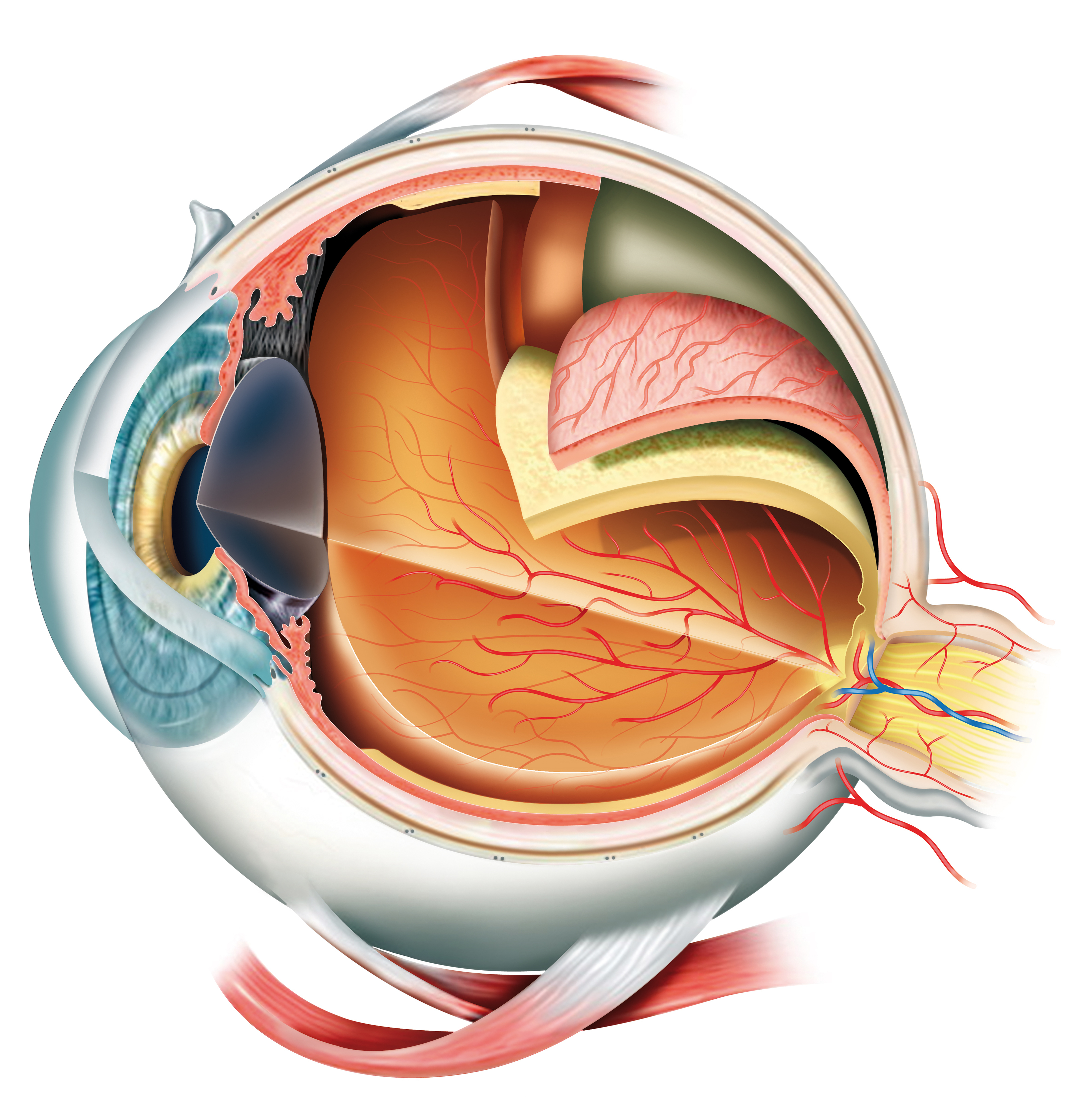
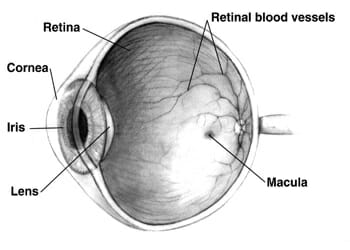
Inner Eye Anatomy
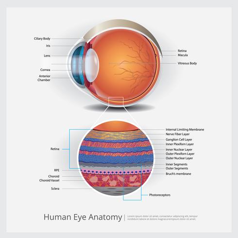
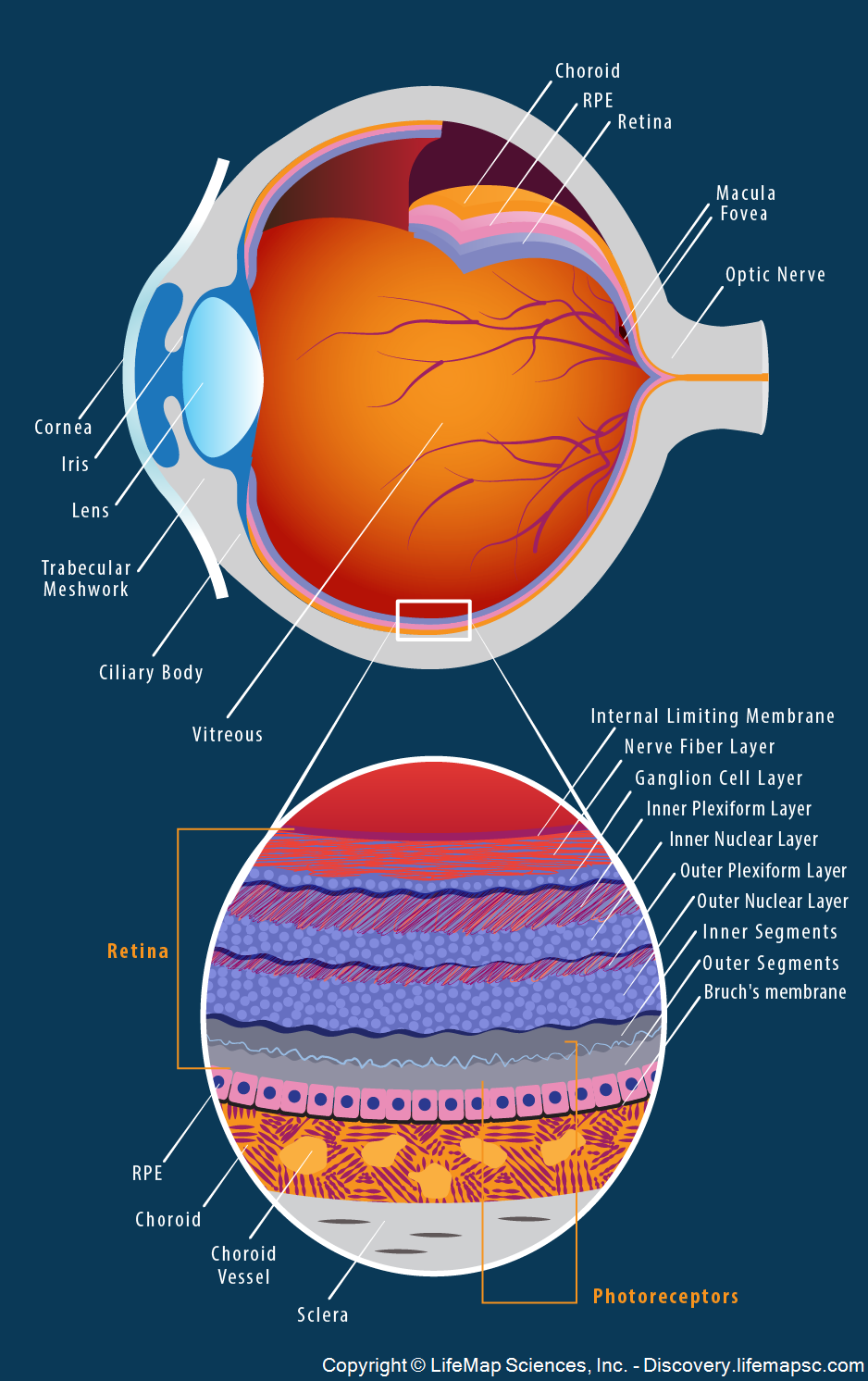
A thickened mass usually on the inner part of your eyeball. The retina itself is composed of two cellular layers.
 The Eye Ear Special Sense Organs Junqueira S Basic
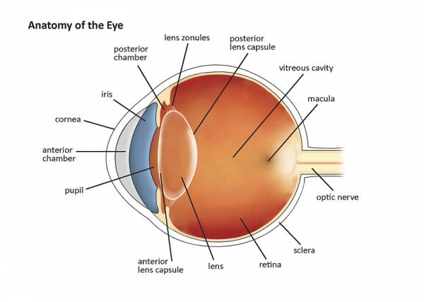
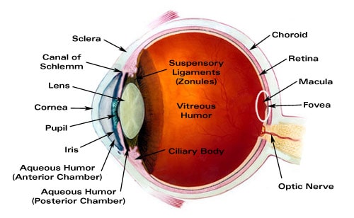
The zonula threads are then attached to the ciliary body.

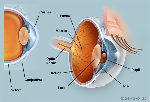
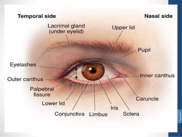
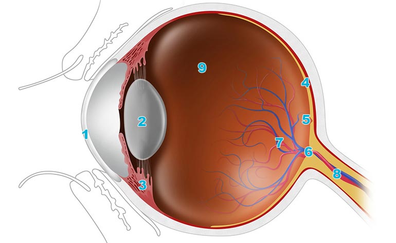
Inner eye anatomy. The conjunctiva is a thin transparent layer of tissues covering the front of the eye. Picture of eye anatomy detail cornea. Deposits of yellowish extra cellular waste products that accumulate within and beneath the retinal pigmented epithelium rpe layer.
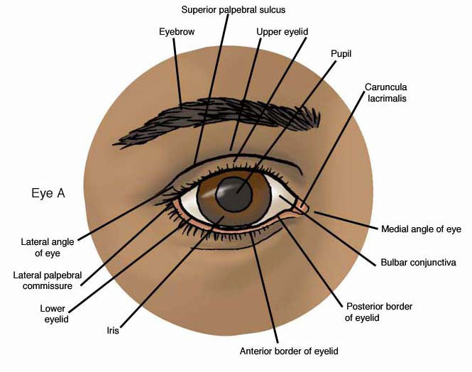
Circular and radial muscle. A closer look at the parts of the eye by liz segre when surveyed about the five senses sight hearing taste smell and touch people consistently report that their eyesight is the mode of perception they value and fear losing most. Colored part of the eye that helps regulate the amount of light that enters.
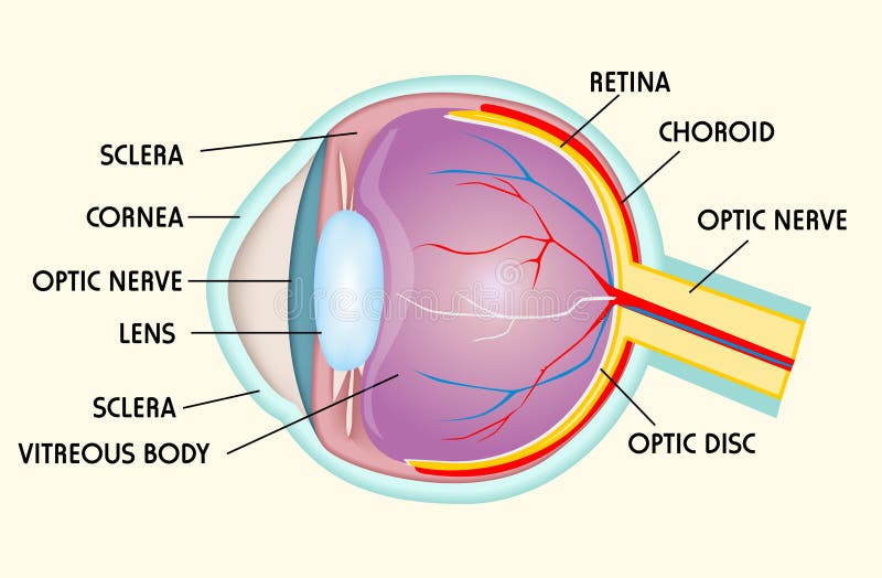
When we then want to focus on a near object a muscle in the ciliary body contracts. The cornea is the transparent clear layer at the front and. Anatomy of the eye.
The outer transparent structure at the front of the eye that covers the iris pupil and anterior chamber. It has small circular opening called pupil. It can cover a part of the cornea and lead to vision problems.
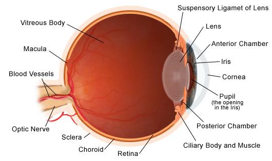
The anatomy of the eye includes the cornea pupil lens sclera conjunctiva and more. The white part of the eye that one sees when looking at oneself in the mirror is. It is muscular pigmented and opaque diaphragm which hangs in the eye ball in front of lens.
The light detecting cells of the retina. It can cover a part of the cornea and lead to vision problems. The inner layer of the eye consists of the retina the light detecting part of the eye.
It consists of photoreceptors. Neural layer the innermost layer of the retina. Anatomy and physiology of the eye conjunctiva.
There are many parts of the eye. We can compare this optic correlation with a bicycle wheel where the lens is the hub the threads the spokes and the ciliary body the rim. It has two types of muscles.
Dark aperture in the iris that determines how much light is let into the eye. It is the eyes primary light focusing structure. Clear front window of the eye that transmits and focuses light into the eye.
 Eye Anatomy Illustration 92433742 Retina Group Of New York
 The Eyes Human Anatomy Diagram Optic Nerve Iris Cornea

 Anatomy Of A Normal Human Eye Amdf
Parts Of The Eye American Academy Of Ophthalmology

 Anatomy Of The Eye Moorfields Eye Hospital
 Anatomy Of Eye
 Stock Illustration
Eye Anatomy Model
 Human Eye Anatomy Vector Illustration Download Free
 Amazon Com Vanfan Durable Shower Curtains Eye Anatomy Inner
 Your Eyes For Kids Nemours Kidshealth
 Class Eye Structure Online Presentation
 Eye Structure Stock Vector Illustration Of Learn Cornea
 The Eye Structure External Parts Of The Eye Tear Duct
 Anatomy Atlases Anatomy Of First Aid A Case Study Approach
 Cross Section Of Human Eye Medical Aid Banner Stock Vector
 Dry Eye
 Structure And Function Of The Eyes Eye Disorders Merck
 Eye Anatomy Glaucoma Research Foundation
 Stock Illustration
 An Easy Guide To Your Eye S Anatomy Lenstore Co Uk
 Blind Spot Anatomy Britannica
 Eye Anatomy Detail Picture Image On Medicinenet Com
 The Eye And Vision
 Eyes Anatomy Overview Parts And Functions Biology
Belum ada Komentar untuk "Inner Eye Anatomy"
Posting Komentar