Mcl Anatomy
It is on the medial inner side of the knee joint in humans and other primates. They are cause by either a direct blow more severe tear or a non contact injury less severe.
 Medial Collateral Ligament An Overview Sciencedirect Topics
Most mcl injuries can be managed conservatively with good results.
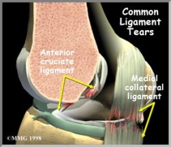
Mcl anatomy. However a complete understanding of knee anatomy and the involved structures is necessary to make intelligent treatment decisions. The medial tibial collateral ligament mcl of the knee is a flat triangular band on its medial aspect and has superficial and deep portions. The medial collateral ligament mcl is the most commonly injured ligament of the knee.
The mcl connects the top of the tibia or shinbone to the bottom of the femur or thighbone. The mcl originates from a sulcus on the distal medial. Medial collateral ligament injury of the knee mcl tear are the most common ligament injuries of the knee and are frequently associated with acl tears.
Medial collateral ligament mcl injury is one of the most common knee injuries especially in young athletic patients. The mcl medial collateral ligament is a band of tissue that runs along the inner edge of your knee. Ligaments hold bones together and add stability and strength to a joint.
The medial collateral ligament is recognised as being a primary static stabiliser of the knee and assists in passively stabilising the joint. When stress is applied this ligament aids control in transferring the joint through a normal range of movement. Gross anatomy it originates at the medial femoral epicondyle anteroinferior to the adductor tuberc.
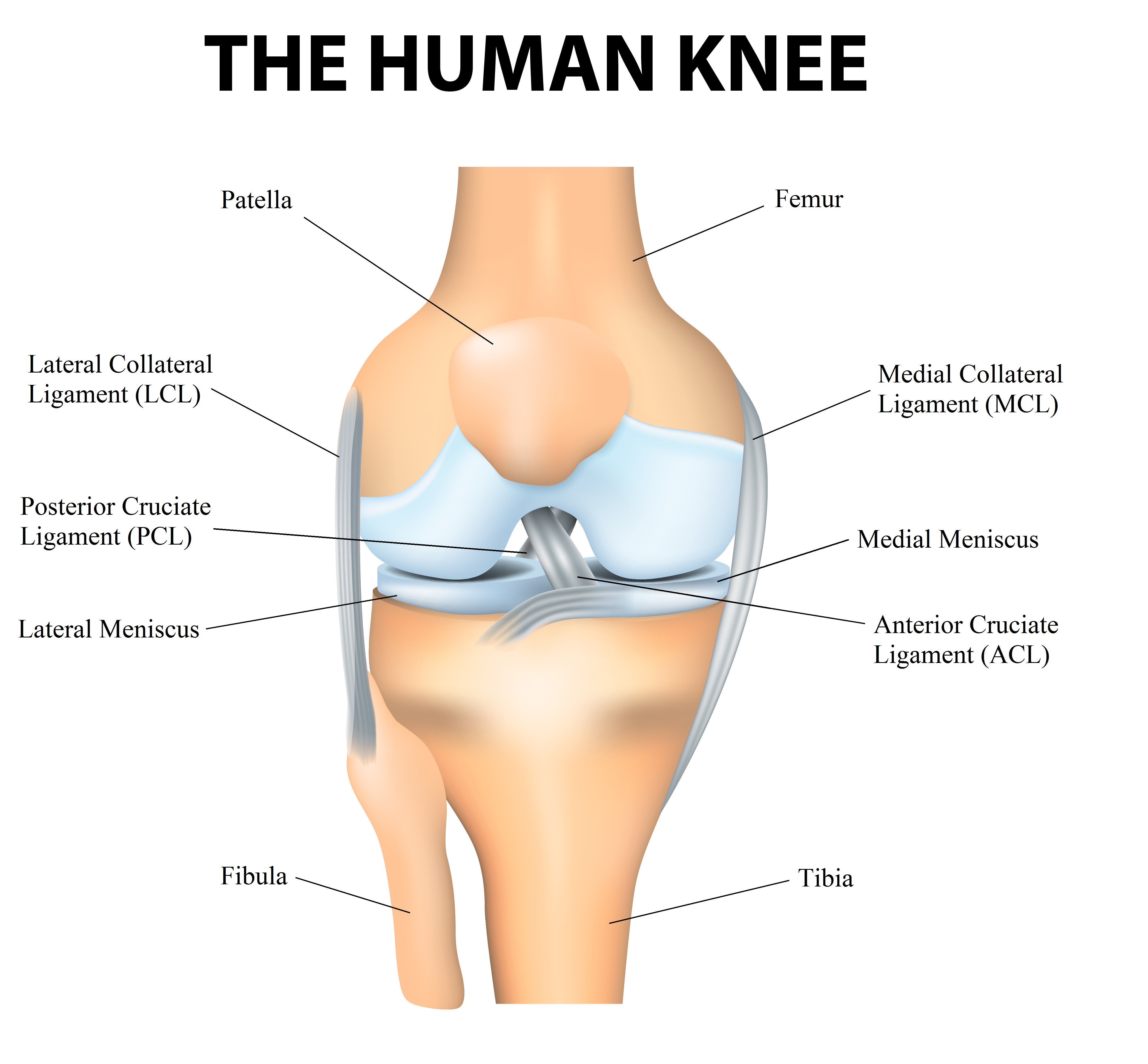
An injury to the mcl is often called an mcl sprain. The mcl also prevents an anterior movement of the tibia and hyperextension. The medial collateral ligament mcl or tibial collateral ligament tcl is one of the four major ligaments of the knee.
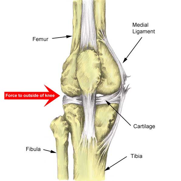
The medial collateral ligament mcl is located on the inner aspect or part of your knee but its outside the joint itself. It helps to connect your shin and thigh bones to keep your knee stable and working properly. The mcl is a strong ligament and it fails in valgus.
Mcl injuries are a common occurrence in sports which require sharp cutting and changing directions and in contact sports. Its primary function is to resist outward turning forces on the knee. The anatomy of the medial side of the knee is pretty intricate and composed of the static stabilizers such as the s mcl d mcl posterior oblique ligament and the posteromedial capsule.
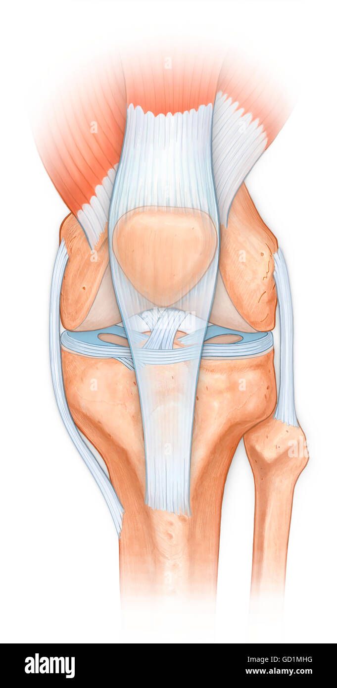
 Pcl Acl Lcl Mcl Meniscus Anatomy
 Knee Joint Ligament Anatomy
 Mcl Tear Of The Knee Injury Diagnosis Treatment
 Medial Collateral Ligament Mcl Radiology Key
 Mcl Injury Active Care Physiotherapy Clinic
:format(png)/cdn.vox-cdn.com/imported_assets/1165294/Gray348.png) A Doctor Explains Dan Henderson S Mcl Tear Bloody Elbow
A Doctor Explains Dan Henderson S Mcl Tear Bloody Elbow
 Medial Collateral Ligament Wikipedia
 6 Illustrations Of The Anatomy Of The Medial Collateral
 Mcl Sprains Part 1 Anatomy Function Mechanism Of Injury
 The Anatomy Of The Mcl
 Mcl Tear Symptoms Diagnosis And Treatment

 Medial Collateral Ligament An Overview Sciencedirect Topics
 Mcl Reconstruction Tendon Injuries Ligament Injuries
 Normal Anatomy Of The Knee Joint Patellar Ligament
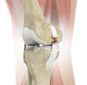 Collateral Ligament Tear Reconstruction Carlsbad Ca Mcl

 Mcl Injury Medial Collateral Ligament Knee Doctor In
 Physical Therapy In Harvard For Knee Collateral Ligament
 Mcl Tears Mark W Maffet M D
Knee Pain On The Inside Of Your Joint Causes Solutions
 Mcl And Lcl Tears
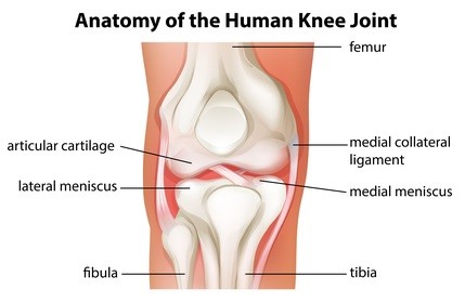
 Knee Anatomy
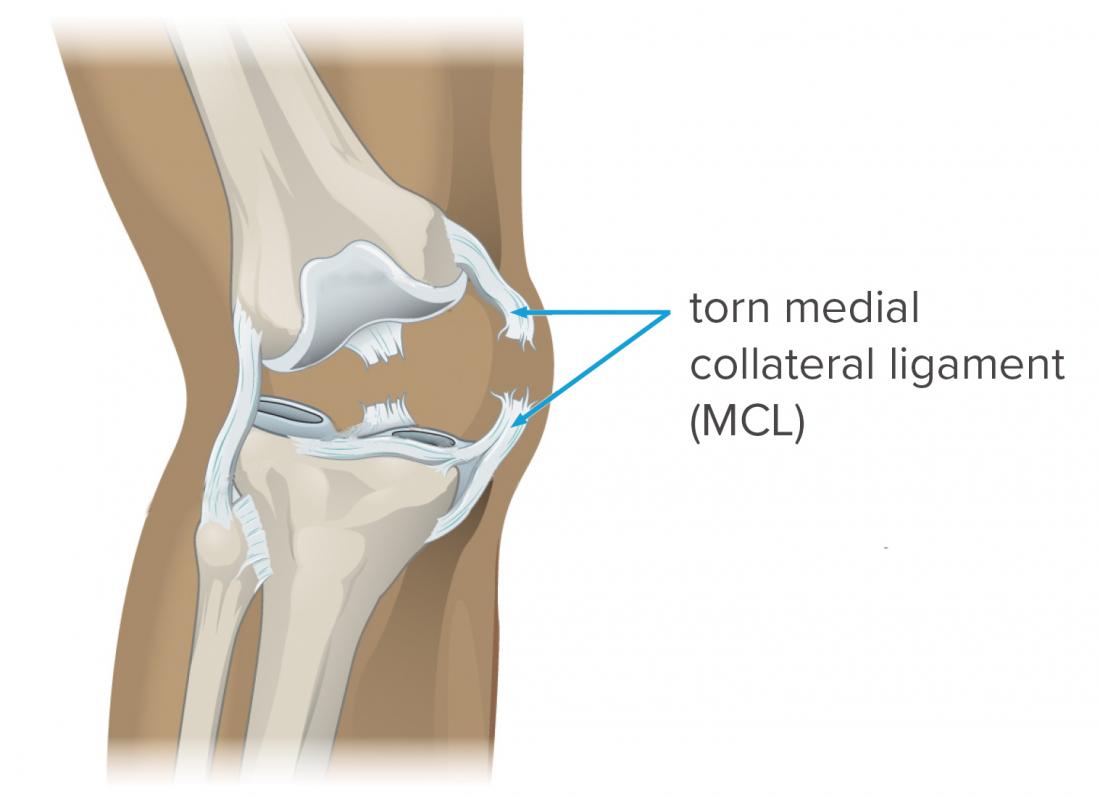
 Do You Have A Torn Mcl
 Medial Collateral Ligament An Overview Sciencedirect Topics
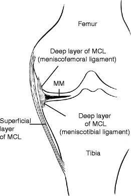
 Deep And Superficial Mcl And Acl Double Bundle Anatomy
 Mcl Injury Torn Medial Collateral Ligament Information
 Medial Knee Ligament Mcl Sprain Symptoms Treatment

Belum ada Komentar untuk "Mcl Anatomy"
Posting Komentar