Svc Anatomy
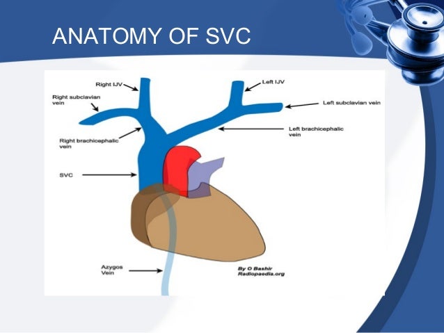
The superior vena cava svc is a large valveless vein that conveys venous blood from the upper half of the body and returns it to the right atrium. Superior vena cava bcv.
 Superior Vena Cava Cardiovascular System Human Anatomy Kenhub
Containing over 1000 vibrant full colour images teachmeanatomy is a comprehensive anatomy encyclopaedia presented in a visually appealing easy to read format.

Svc anatomy. The lung consists of five lobes. The superior vena cava svc is the superior of the two venae cavae the great venous trunks that return deoxygenated blood from the systemic circulation to the right atrium of the heart. Diagnosis not applicable diagnosis not applicable.
Created by a team of doctors and medical students each topic combines anatomical knowledge with high yield clinical pearls seamlessly bridging the gap between scholarly learning and. The superior vena cava svc also known as the cava or cva is a short but large diameter vein located in the anterior right superior mediastinum. Whereas many mammals including humans have only one anterior vena cava other animals have two.
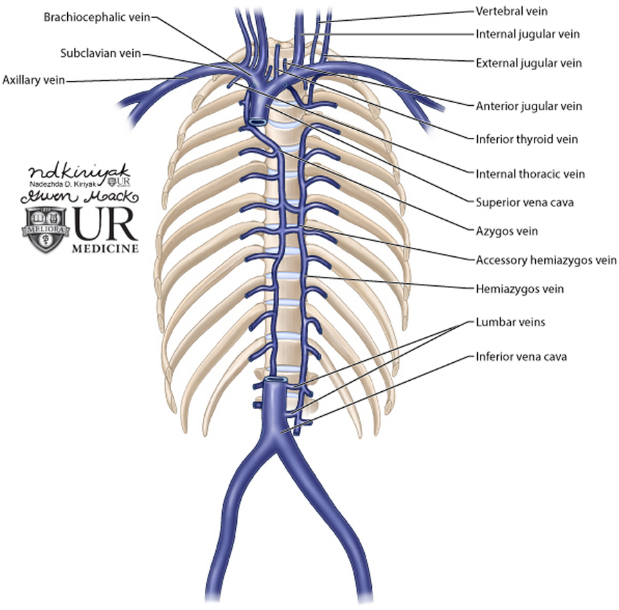
In humans these veins are respectively called the superior and inferior venae cavae. It is a large diameter 24 mm short length vein that receives venous return from the upper half of the body above the diaphragm. The left lung has a superior and inferior lobe while the right lung has superior middle and inferior lobes.
The anterior vena cava also known as the precava drains the head end of the body while the posterior vena cava or postcava drains the tail or rear end. Clinical notes superior vena cava obstruction svco superior vena cava syndrome svcs superior vena cava thrombosis. The superior vena cava svc is a large valveless venous channel formed by the union of the brachiocephalic veins.
Gross anatomy the svc be. Superior vena cava bcv. Case contributed by dr omar bashir.
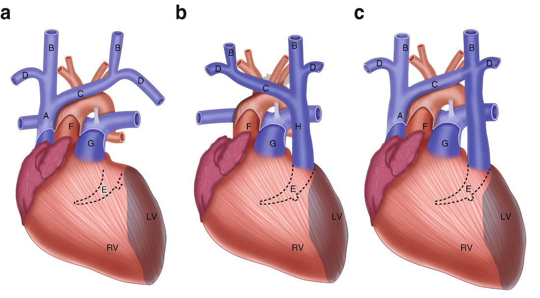
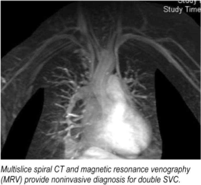
Superior vena cavaon left in anatomy a persistent left superior vena cava plsvc is the most common variation of the thoracic venous system 1 2 is prevalent in 03 of the population 3 and an embryologic remnant that results from a failure to involute. In this article we will look at the anatomy of the superior vena cava its position tributaries and clinical correlations. Thin walls of tissue called fissures separate the.
It receives blood from the upper half of the body except the heart and returns it to the right atrium.
 Persistent Left Superior Vena Cava Springerlink
 1000 Superior Vena Cava Stock Images Photos Vectors
 Learnoncology
 Supra Vena Cava Obstruction Svco
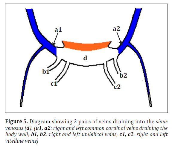
 Sinus Venosus An Overview Sciencedirect Topics
 Cardiology Anatomy Embryology Flashcards Quizlet
 Left Superior Vena Cava With Associated Venous Variations
![]() Superior Vena Cava Anatomy Function Clinical Aspects
Superior Vena Cava Anatomy Function Clinical Aspects
Persistent Left Svc Absent Innominate Bridging Vein
 Shrapnel In The Svc Inset Shrapnel Download Scientific
 Cavoatrial Junction Wikipedia

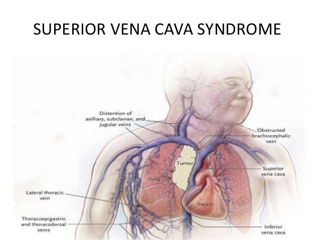 Superior Vena Cava Syndrome
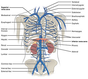 Brachiocephalic Vein Wikipedia


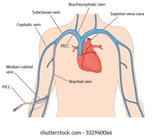
 Misplaced Central Venous Catheters Applied Anatomy And
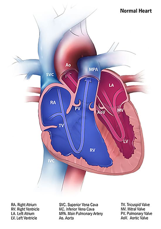 Congenital Heart Defects How The Heart Works Cdc
 Superior Vena Cava And The Azygos System Clinical Anatomy Svc Obstruction Oncology Emergency
 Superior Vena Cava Syndrome Pancoast Syndrome
 Endovascular Occlusion Balloon For Treatment Of Superior
 Sinus Venosus An Overview Sciencedirect Topics
![]() Superior Vena Cava Anatomy Function Clinical Aspects
Superior Vena Cava Anatomy Function Clinical Aspects
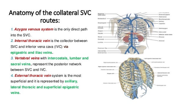
Where There Is Blood There Is A Way Unusual Collateral

 Supra Vena Cava Obstruction Svco
 Superior Vena Cava An Overview Sciencedirect Topics
 Blood Finds A Way Pictorial Review Of Thoracic Collateral
 Cureus A Route Less Traveled Anomalous Venous Drainage Of
 Superior Vena Cava Syndrome Occurs When The Svc Is
 Learnoncology
 Atrium Superior Vena Cava Svc Inferior Vena Cava Ivc And
 Easy Notes On Superior Vena Cava Learn In Just 3 Minutes
Belum ada Komentar untuk "Svc Anatomy"
Posting Komentar