Gastrocnemius Vein Anatomy
They are embedded in the belly of calf muscles such as the soleus and gastrocnemius and are able to dilate and hold a large amount of blood. Sat outside for hours in short lil chilly yesterday started.
Venous sinuses are closely related to deep veins.
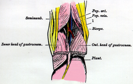
Gastrocnemius vein anatomy. The gastrocnemius is a powerful plantar flexor of the foot at the talocrural joint. In one cadaver based series the number of gastrocnemius veins varied from 2 12 for each muscle head. The main gastrocnemius venous trunks were opened longitudinally to allow assessment of the presence and number of valves.
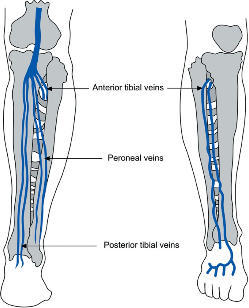
The anatomy of the gastrocnemius and soleus veins is variable. The posterior tibial anterior tibial and peroneal veins lie between the muscles. Place the probe transversely at the knee crease in the popliteal fossa.
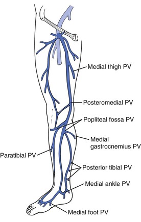
It also flexes the leg at the knee. The medial and lateral gastrocnemius veins which drain the intramuscular zone. And finally the popliteal vein sometimes duplicated by a collateral vessel which drains blood from the intermuscular.
Feels pain in touch. 3a most of the gastrocnemius veins drained into the popliteal vein fig. Check the compressibility of the popliteal vein throughout the popliteal fossa.
Locate the popliteal artery and vein. All gastrocnemius veins were dissected from the gastrocnemius muscle heads toward their drainage site. The anatomy of the iliac veins has been less thoroughly described than that of the infrainguinal veins.
The ability to visualize the gastrocnemius veins with mr is variable due in part the compressibility of the veins. The deep vein extending from the popliteal vein to the common femoral vein is now referred to as the femoral vein rather than the superficial femoral vein. The soleal sinuses and gastrocnemius veins lie within the muscles.
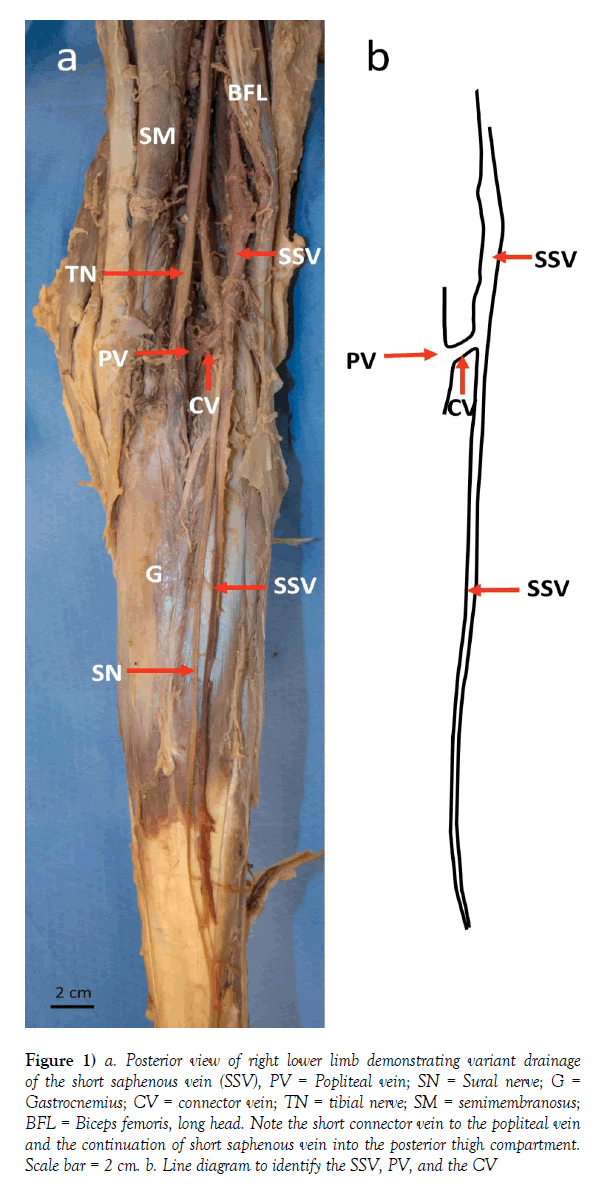
Venous drainage is through corresponding medial and lateral sural veins into the popliteal vein. Be cautious not to mistake the often prominant muscular veins gastrocnemius veins for the popliteal vein. The actions of gastrocnemius are usually considered along with soleus as the triceps surae group.
The gastrocnemius muscle plural gastrocnemii is a superficial two headed muscle that is in the back part of the lower leg of humans. The short saphenous vein and its collaterals which drain the subcutaneous zone. The popliteal fossa a real venous crossroads is the site of anastomosis of three superimposed venous planes.
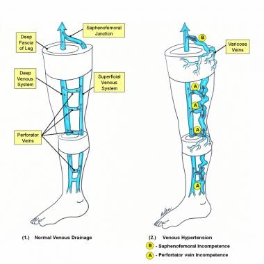
With the contraction of calf muscles at walking the blood is pumped to more proximal deep veins calf muscle pump. All the deep veins of the calf join to form the popliteal vein which is the calf pump outflow tract. It runs from its two heads just above the knee to the heel a three joint muscle knee ankle and subtalar joints.
Gastrocnemius has a red vein visible from the skin.
Should Cardiologists Be Involved In The Management Of
 Module 2 Lower Extremity Orthopedic Imaging
 Varicosities Affecting The Lower Limb Veins Consequent To A
 Figure 1 From Is The Treatment Of The Small Saphenous Veins
 Gastrocnemius Muscle Knee Low Leg Ankle Arch Pain The
 Varicose Vein Surgery Practice Essentials Anatomy
 Popliteal Fossa And Knee Joint Last S Anatomy Regional
 Ecr 2014 C 1493 Practical Approach To Varicose Veins In
 Venous Anatomy Plastic Surgery Key
 Anatomy Atlases Illustrated Encyclopedia Of Human Anatomic
 Doppler Ultrasound In Deep Vein Thrombosis
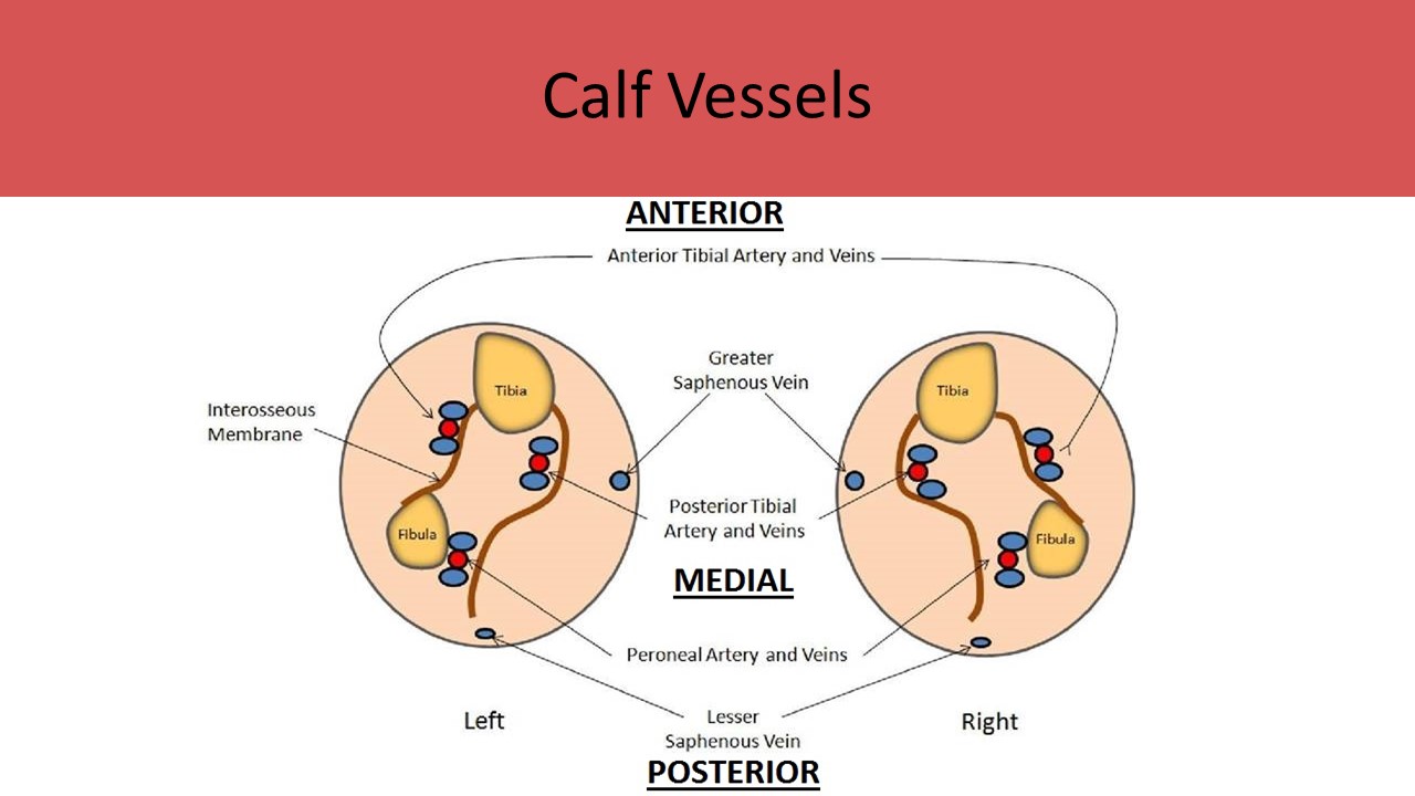 Ultrasound Registry Review Extremity Venous
 The Venous System Of The Foot Anatomy Physiology And
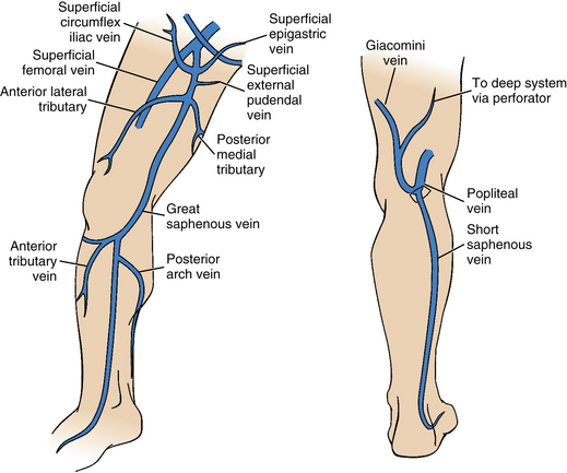 Lower Extremity Veins Radiology Key
Popliteal Artery Entrapment A Mysterious Syndrome
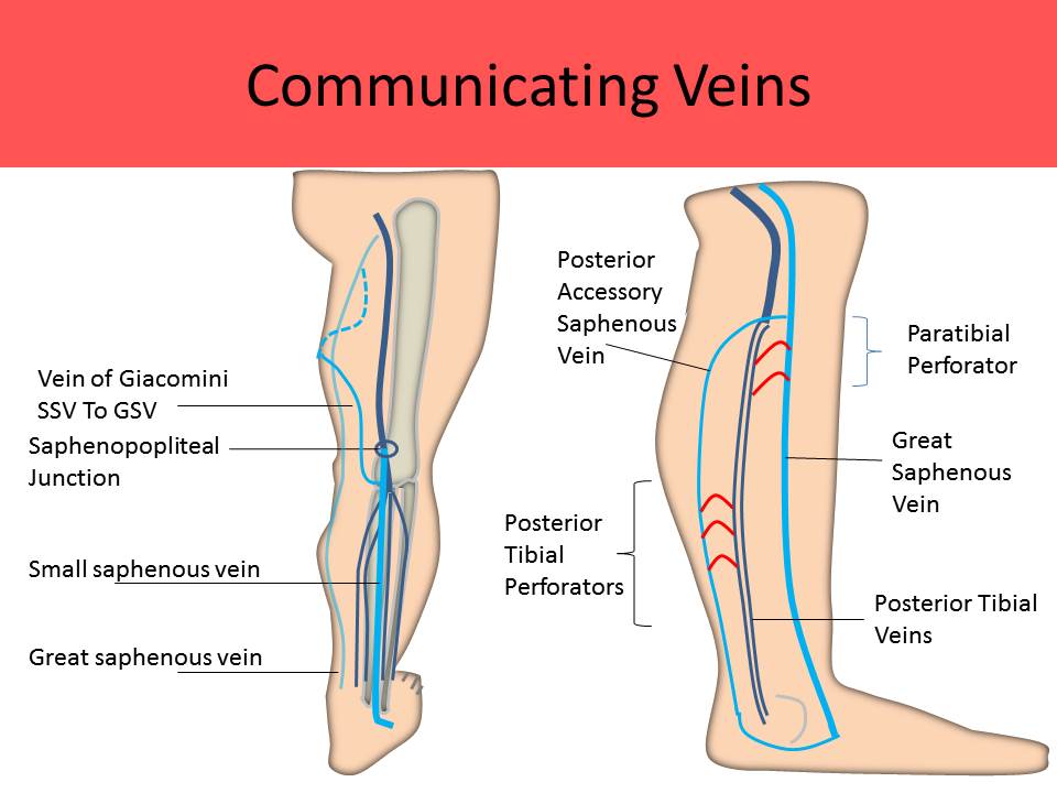 Ultrasound Registry Review Extremity Venous
 Ectatic Gastrocnemius Veins Phlebologia
Gastrocnemius Vein Anatomy Elegant Evidence For Treatment Of
 Popliteal Vein Radiology Reference Article Radiopaedia Org
 Difference In The Anatomical Structure Between The



Belum ada Komentar untuk "Gastrocnemius Vein Anatomy"
Posting Komentar