Anatomy Of Vision
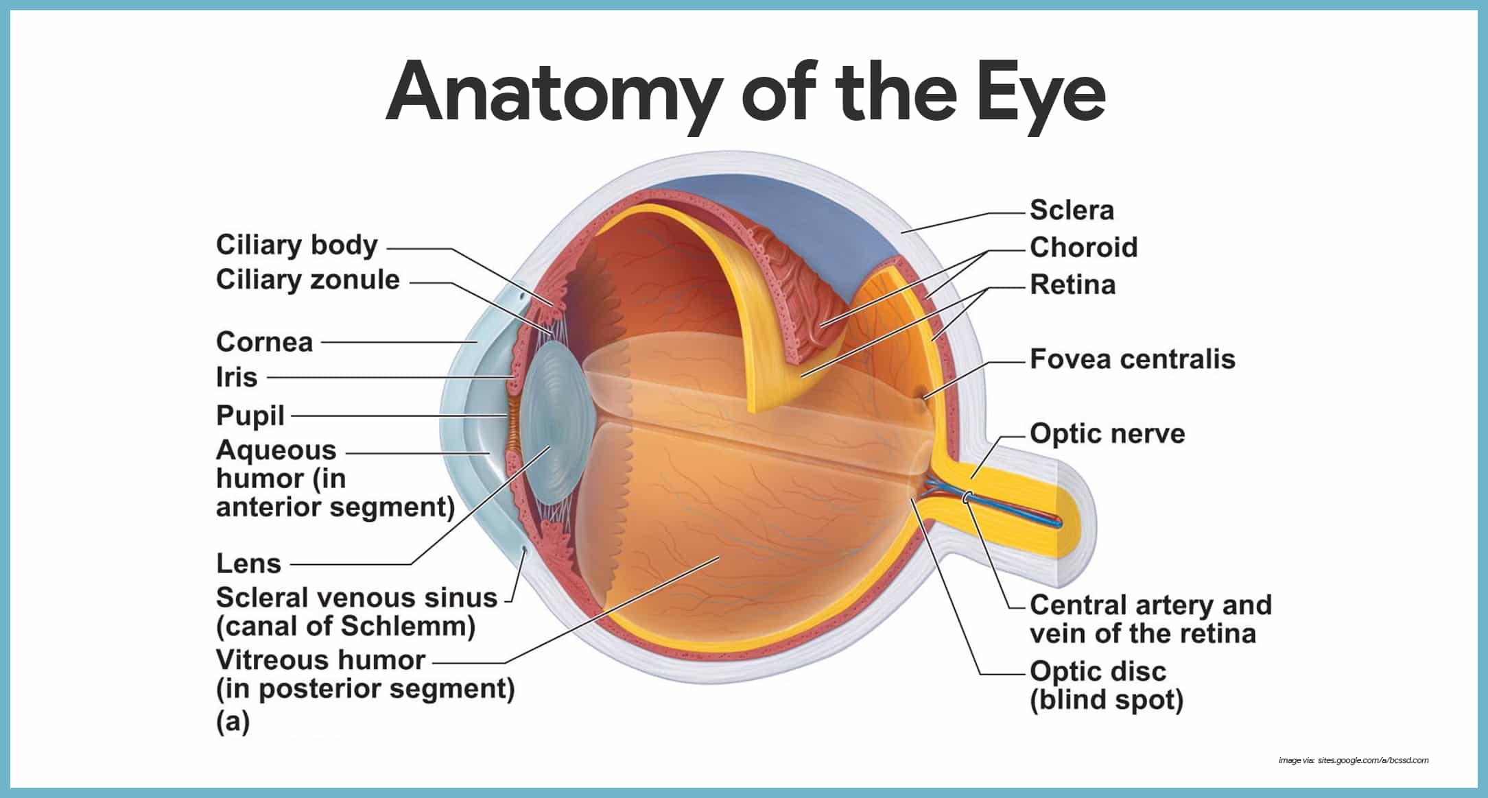
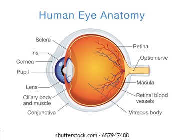
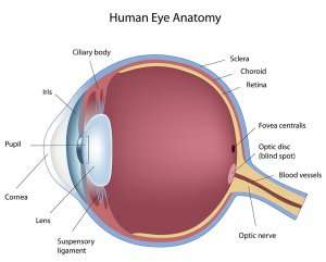
The iris has two muscles. The color of the iris is determined by the color of the connective tissue and pigment cells.
 Vision Lab Review Eye Anatomy Especially Ppt Download
Four of the muscles are arranged at the cardinal points around the eye and are named for those locations.

Anatomy of vision. Less pigment makes the eyes blue. The dilator muscle makes the iris smaller and therefore the pupil larger. Eyes are beautiful thingsthey must be.
Image from human anatomy atlas. Image from human anatomy atlas. The iris of the eye functions like the diaphragm of a camera controlling the amount.
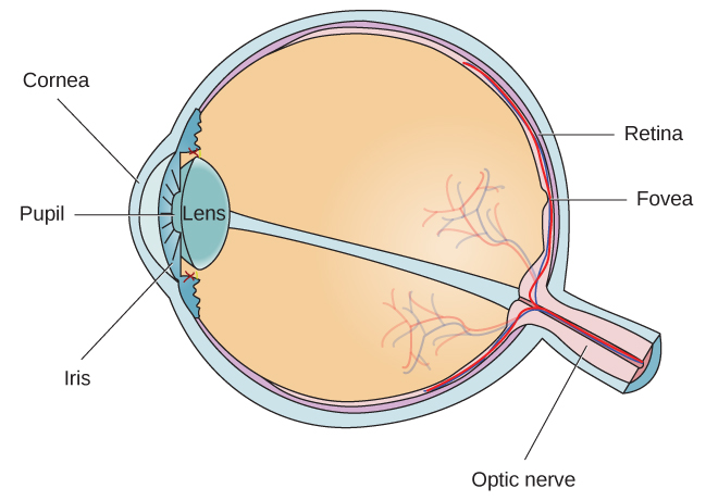
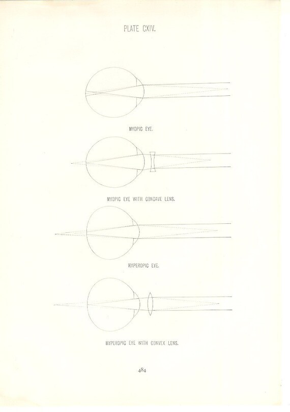
The cornea is the transparent clear layer at the front and. The conjunctiva is a thin transparent layer of tissues covering the front of the eye. How vision works sight begins when light rays from an object enter the eye through the cornea the clear front window of the eyeball.
Anatomy and physiology of the eye conjunctiva. Movement of the eye within the orbit is accomplished by the contraction of six extraocular muscles that originate from the bones of the orbit and insert into the surface of the eyeball figure 2. Light is focused primarily by the cornea the clear front surface of the eye.
Optic nerve and. In a number of ways the human eye works much like a digital camera. The white part of the eye that one sees when looking at oneself in the mirror is.
The iris is an adjustable diaphragm around an opening called the pupil. More pigment makes the eyes brown. The eyes crystalline lens is located directly behind the.
The cornea is actually responsible for about sixty percent of the eyeballs light ray bending capability. The anatomy of vision orbits eyesockets like most structures in the body the orbits of the skull do more than one job.
 Special Senses Vision Anatomy And Physiology I
 Vision And The Eye S Anatomy Healthengine Blog
 Hand Drawn Human Eye With Iris Anatomy Of Vision Organ
 4d Vision Brick Man Anatomy Model

 The Anatomy Of Vision A Section Through Eye Showing The
 Cataracts Vision Disorder And Normal Eye Vision Anatomy Vector
 Special Senses Anatomy And Physiology Nurseslabs
 I Can See Clearly Now The Tumor S Gone Pacific
 Vision Introduction To Psychology
 Retinopathy Is Damage To The Retina Of The Eyes Which Cause
 Human Eye Cross Section Normal Vision Stock Vector

Eye Anatomy Glaucoma Research Foundation
 Hand Drawn Human Eye With Iris Anatomy Of Vision Organ
 Eye Anatomy Vision Images Stock Photos Vectors Shutterstock
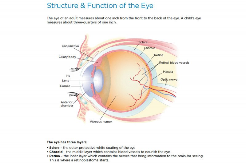 Retinoblastoma Anatomy Of The Eye Memorial Sloan
 Device Renews Hope For Artificial Vision Ophthalmology
 How Vision Works Eye Science Projects Experiments Hst
 How Your Retina Works How Artificial Vision Will Work
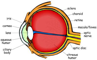
 Eye Anatomy Ozarks Family Vision Centre
 Sight Br Eye And Vision Illustration Of The Anatomy Of The
 Eye Anatomy Structure Function Of Vision Hs Ls1 A By
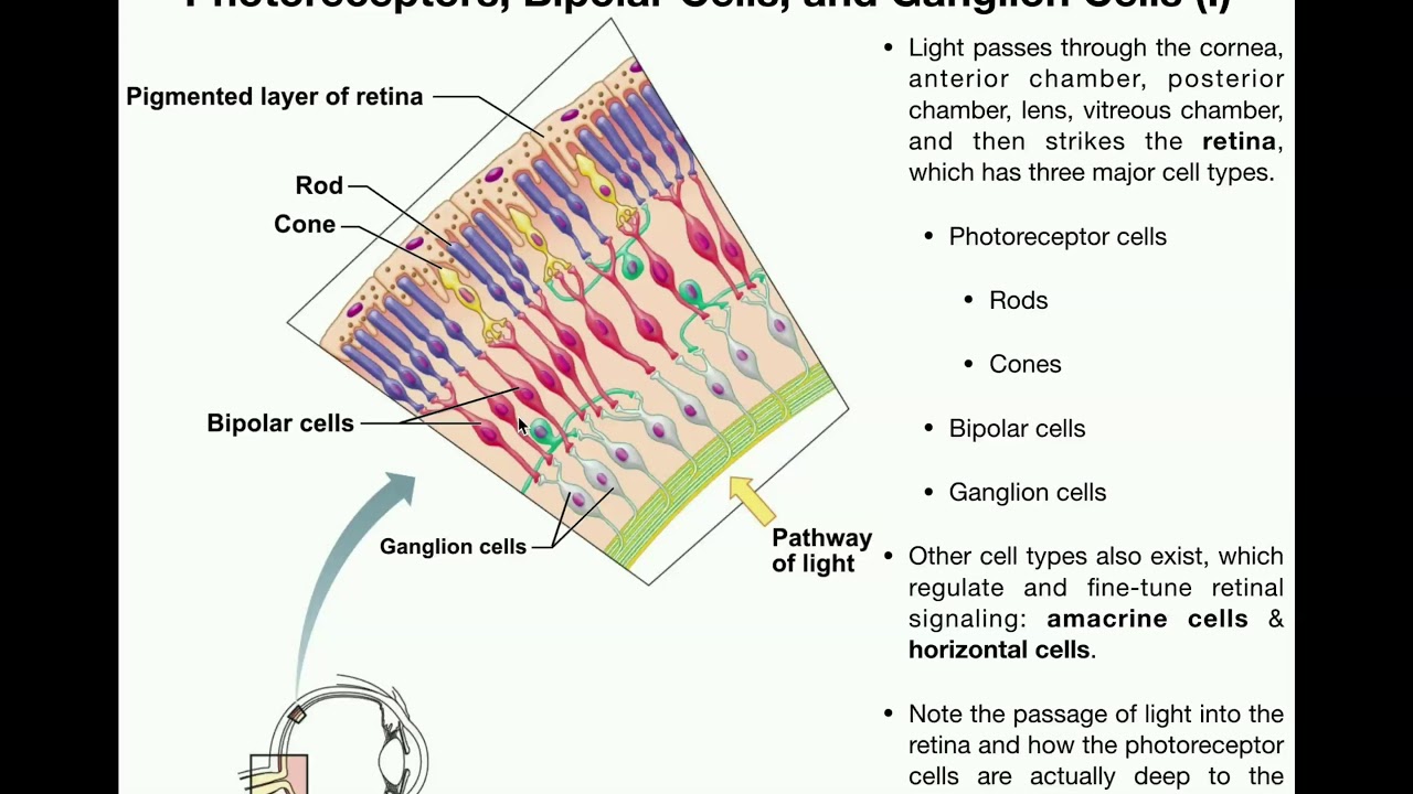 Anatomy Vision Part 1 Retina Photoreceptors Bipolar Cells Ganglion Cells
 4d Vision Human Torso Anatomy Model

 Anatomy 1926 Human Anatomy Print Vision Eye Chart Vintage Antique Medical Anatomy Art Illustration For Doctor Hospital Office
 Eye Anatomy 101 American Eyecare Optometrist In Keokuk Ia
 Pin On Pca Effects The Back Of The Brain
Belum ada Komentar untuk "Anatomy Of Vision"
Posting Komentar