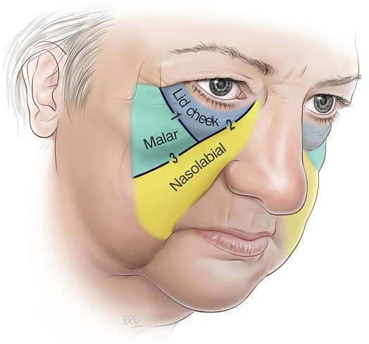
Face Surface Anatomy
It marks the position of the stylomastoid foramen which lies about 2 cm deep to the surface in adult. Surface anatomy of the face and skin.
Illustration of the human face labelled.

Face surface anatomy. Surface anatomyface 2. They serve however to round off and smooth prominent borders and to fill up what would otherwise be unsightly angular depressions. The outlines of the muscles of the head and face cannot be traced on the surface except in the case of the masseter and temporalis.
4446 gbp why the license you choose matters. The fibres are ordered into upper intermediate and lower groups. Cranium can be subdivided into three regions each having prominent surface anatomy features.
Roll mouse over image to display labels. Those of the face are small covered by soft skin and often by a considerable layer of fat and their outlines are therefore concealed. This application allows for the precise and comprehensive labeling of anatomic locations of dermatologic disease thereby reducing biopsy and treatment site ambiguity and providing a rich dataset upon which data mining can be performed.
Head surface anatomy 1. Medical gross anatomy surface anatomy. Start studying surface anatomy of the face.
The muscles of the scalp are so thin that the outline of the bone is perceptible beneath them. The surface anatomy of the face is best appreciated by referring to the bony landmarks of the frontal maxillary zygomatic and mandibular bones. Each of these bones has distinctive features that contribute to the surface landmarks of the facethe orbital rims zygomatic arch the mastoid process and the mentum.
A web based human surface anatomy mapper was developed to allow labeling of mapped surface anatomy images. A point on the middle of the anterior border of the mastoid process. Home surface anatomy of the face and skin.
Learn vocabulary terms and more with flashcards games and other study tools. Caucasianprominent nose narrow nasal aperture big brow ridges large jaws and teeth prognathous. 13 6 cranium cranium cranial region or braincase is covered by the scalp which is composed of skin and subcutaneous tissue.
The frontal region of the cranium is the forehead covering the frontal region is the frontalis muscle which overlies the frontal bone the frontal region terminates at the superciliary arches. Converging muscles around the mouth after origin the fibres run in the direction of the mouth and fill the gap between the upper and lower jaws. On surface projection the cranial exit of the nerve lies just in front of the intertragic notch grays anatomy38th ed.
 Surface Anatomy Of The Face And Skin
 Surface Anatomy Head Neck Surface Anatomy A Branch Of
 Head Surface Anatomy 4 Edition
 The Double Face Of The Periorbital Zone English French
 Paranasal Sinus Anatomy Overview Gross Anatomy
 Human Surface Anatomy Labeling System Matt Molenda Md
 Colorful Surface Anatomy Of Face Ornament Internal Organs
 Section 8 Atlas Of Surface Anatomy Hadzic S Peripheral
 Surface Anatomy The Medical Textbook
 Surface Anatomy An Overview Sciencedirect Topics
 World S Best Human Head Stock Illustrations Getty Images
 Facelift Anatomy Smas Retaining Ligaments And Facial
 Surface Anatomy Biology 3418 With Dr Mcnair At Hardin
 The Face Pictorial Atlas Of Clinical Anatomy 9781850972907
 Surface Anatomy An Overview Sciencedirect Topics
 Surface Markings Of Special Regions Of The Head And Neck
 Useful Notes On The Surface Anatomy Of Head And Neck Human
 Surface Anatomy Biology 3418 With Dr Mcnair At Hardin
 Nasal Surface Anatomy Facial Anatomy Face Anatomy Skin
 Head Surface Anatomy 4 Edition
 Chapter 1 2 Intro Surface Anatomy At Florida Southwestern
 List Of Human Anatomical Regions Wikipedia
 Retaining Ligaments Of The Face Review Of Anatomy And
 Anatomy Of Face Wiring Diagram Symbols And Guide
 Anatomy Surface Limbs Limb Lips Woman S Face T Shirts

Belum ada Komentar untuk "Face Surface Anatomy"
Posting Komentar