Mri Ankle Anatomy
37 magnetic resonance imaging mri the ankle is the joint that is located between the leg and the foot. Mr imaging of the ankle and foot introduction.
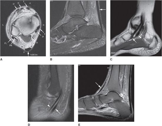
 Acute Spring Ligament Complex Tear Imaging Olympic Park
This module is a comprehensive and affordable learning tool for medical students and residents and especially for physicians anatomists rheumatologists orthopaedic surgeons and radiologists.
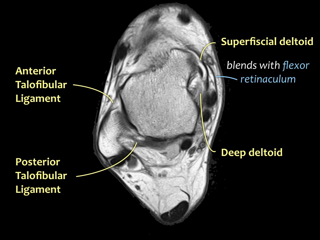
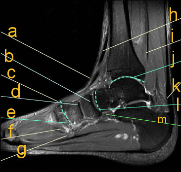
Mri ankle anatomy. On mri the ligaments appear as thin linear low signal intensity structures connecting adjacent bones usually delineated by high signal intensity fat. Start your exam with fatsat images of the bones to screen for edema. Routine ankle mr imaging is performed in the axial coronal.
It demonstrates abnormalities in the bones and soft tissues before they become evident at other imaging modalities. Once you have studied the bones scan the joints for effusion. Use the mouse to scroll or the arrows.
Knee shoulder shoulder arthrogram ankle elbow. Internal derrangements of joints. It is also a fundamental communication tool to teach patients anatomy and pathology.
Mri of the ankle and feet. Racsuq advanced surgical anatomy course upper and lower limbs. Screen on fatsat images for bone marrow edema.
This webpage presents the anatomical structures found on ankle mri. Three ligamentous groups support the ankle joint. This joint is a main contributor of stability in the lower limbs and it allows humans to perform actions such as running jumping and walking 1 2.
Magnetic resonance mr imaging has opened new horizons in the diagnosis and treatment of many musculoskeletal diseases of the ankle and foot. Rmhalf msk ankle. Uq med year 1 gafradiographic anatomy lower limb.
The ligamentous groups that support the ankle joint include the lateral complex the medial complex deltoid ligaments the ankle syndesmosis and the spring calcaneonavicular ligament complex. Ankle mri examination systematic approach. Scroll through the image stack for the.
Mri of the ankle. 663 3 normal extremity. Normal mri ligament anatomy.
The past 15 years have witnessed an explosion of information regarding the role. Click on a link to get sagittal view t1 axial view t2fatsat coronal view t2fatsat sagittal view t2fatsat.
 Leg Mri Stock Photo Download Image Now Istock
 Musculoskeletal Mri
 Imaging Anatomy Interactive Pacs Like Atlas Of Radiological
 Mri Ankle Anatomy
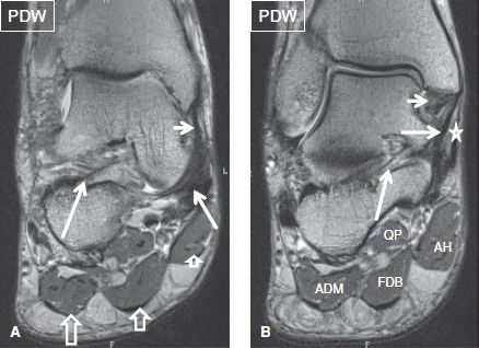
 Mri Of The Ankle Detailed Anatomy
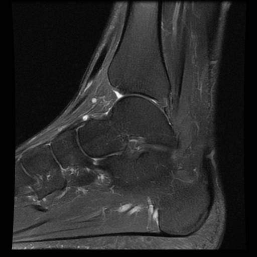
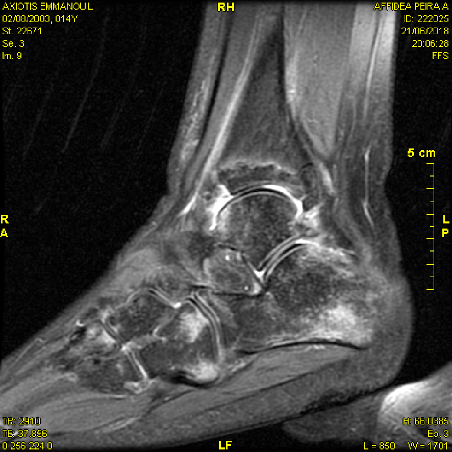
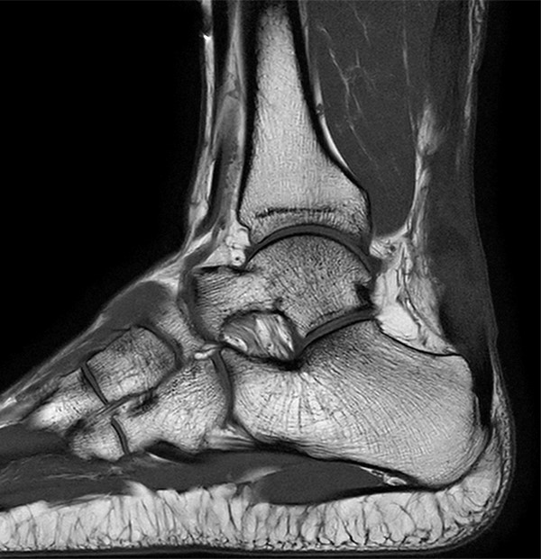
 Mri T2 Image Is Showing Sagittal Section Of The Ankle
 The Radiology Assistant Ankle Mri Examination
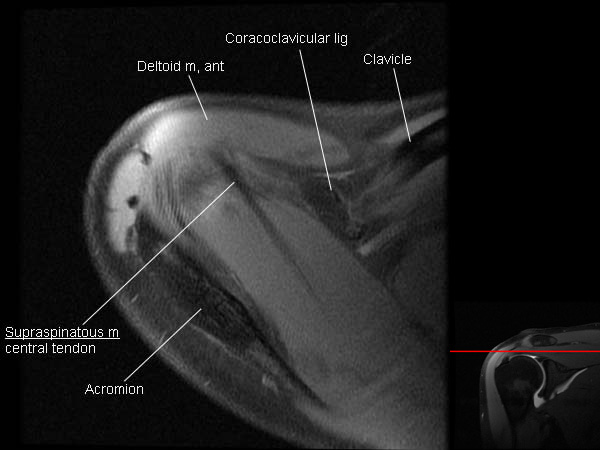 Mri Shoulder Arthrogram Anatomy
 Lower Extremity Os Foot Ankle Orthobullets
 Mri Ankle Anatomy Ankle Anatomy Anatomy Human Anatomy
The Ankle Musculoskeletal Key
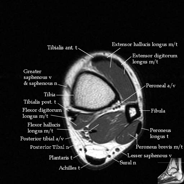
 Mri Anatomy Of Ankle
Mri Imaging Techniques Ankle Deltoid Ligament Injury Mri
 Knee Imaging Knee Sports Orthobullets
 Mri Of The Ankle Detailed Anatomy
 Update On Diagnosis And Management Of Cuboid Fractures
 Ankle Mri Radiology Key
 The Radiology Assistant Ankle Mri Examination
 Mri Imaging Of Soft Tissue Tumours Of The Foot And Ankle
 Ankle Mri Radiology Key
 Courses Mri Online
 The Radiology Assistant Ankle Mri Examination
 Mri Anatomy And Imaging Proprofs Quiz
 Achilles Tendon Pathology Radsource
 High Ankle Sprains Radsource
 Staging Of Osteochondral Lesions Of The Talus Mri And Cone
![]() Stanford Msk Mri Atlas C 2019
Stanford Msk Mri Atlas C 2019


Belum ada Komentar untuk "Mri Ankle Anatomy"
Posting Komentar