Posterior Knee Anatomy
Transition is at hiatus of adductor magnus muscle. The knee functions as a modified hinge joint consisting of the tibia femur and patella.
 Injuries To The Posterolateral Corner Of The Knee Rayner
We are pleased to provide you with the picture named posterior view of knee jointwe hope this picture posterior view of knee joint can help you study and research.
Posterior knee anatomy. Increased posterior translation on the posterior drawer test indicates a combined posterior cruciate ligament tear with the pcl injury. Here we explain the common causes of both sudden onset and chronic overuse pain behind the knee. The posterior cruciate ligament prevents the femur from.
However when pathology is present abduction adduction internal and external rotations and anterior and posterior translations may occur the distal femur and proximal tibia form the two largest points of contact in the. Anchored by insertion of adductor magnus as enters region of posterior knee. Posterior knee pain is a common patient complaint.
Origin before knee a continuation of the superficial femoral artery. Acute posterior knee pain is sudden onset and includes sprains and strains. Knee pain is more common in the anterior medial and lateral aspect of the knee than in the posterior aspect of the knee.
Course in posterior knee relation to anatomy structures of knee lies posterior to the posterior horn of the lateral horn of the lateral meniscus. The posterior and lateral anatomy of the knee joint presents a challenge to even the most experienced knee surgeon. Gradual onset or chronic knee pain develops over time and is often caused by overuse.
Less common are neurologic and vascular injuries. The differential diagnoses for posterior knee pain include pathology to the bones musculotendinous structures ligaments andor to the bursas. Knowledge of the bony topography will result in a greater number of anatomic ligament reconstructions a lack of familiarity leads to hesitancy when performing approaches in these areas of the knee.
Knowledge of the bony topography will result in a greater number of anatomic ligament reconstructions a lack of familiarity leads to hesitancy when performing approaches in these areas of the knee. Knowing which ones typically cause pain in which particular areas of the knee help make it much easier to get an accurate knee pain self diagnosis. A knee pain diagnosis chart can be a really tool to help you work out why you have pain in your knee.
Pain at the back of the knee is known as posterior knee pain. The primary plane of motion is extension and flexion. There are lots of different structures in and around the knee that can cause pain.
Webmds knee anatomy page provides a detailed image and definition of the knee and its parts including ligaments bones and muscles. Figure 4 test the patient lies supine and flexes their affected knee to approximately 90 then crosses it over the normal side with the foot across the knee and the hip externally rotated. The posterior and lateral anatomy of the knee joint presents a challenge to even the most experienced knee surgeon.
For more anatomy content please follow us and visit our website.
 Posterior Cruciate Ligament Reconstruction An Overview
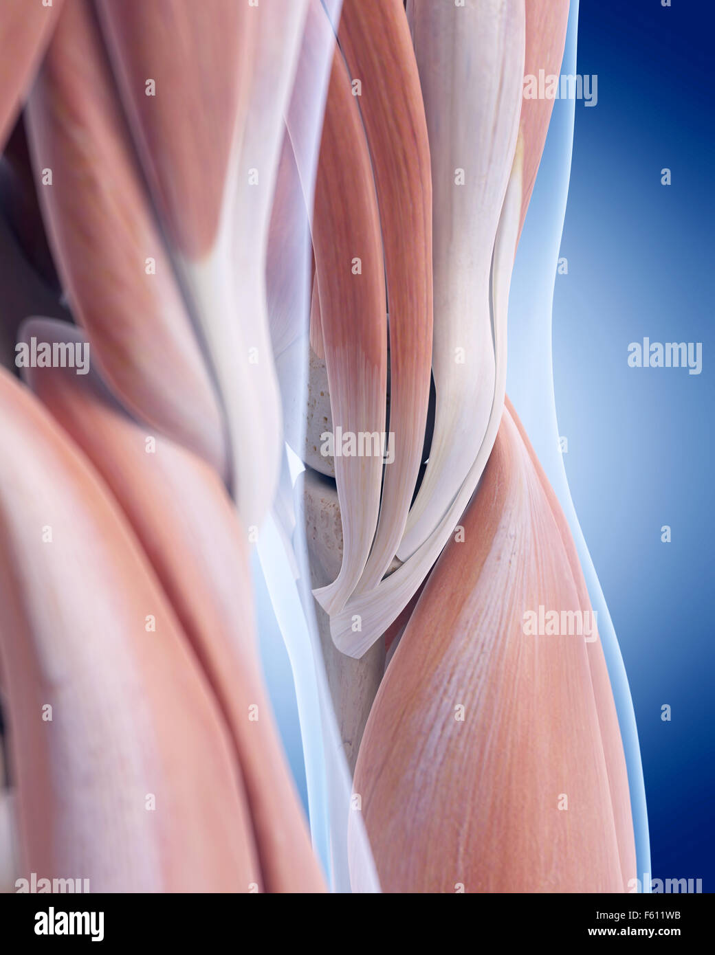 Medically Accurate Illustration Of The Posterior Knee
 Posterior Cruciate Ligament An Overview Sciencedirect Topics
 Knee Joint Picture Image On Medicinenet Com
 Acl Tears Pinnacle Orthopaedics
 Posterior Lateral Corner Injury
 Injuries To The Posterolateral Corner Of The Knee Rayner
 Knee Surgeon Marc Hirner Orthopaedic Surgeon
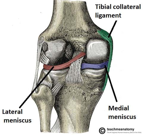 The Knee Joint Articulations Movements Injuries
 Knee Pain In Runners Part 1 A Quick Anatomy Lesson
 Pcl Injury Knee Sports Orthobullets
 Posterior View Of The Right Knee In Extension
 Muscles Of The Leg Part 1 Posterior Compartment Anatomy Tutorial
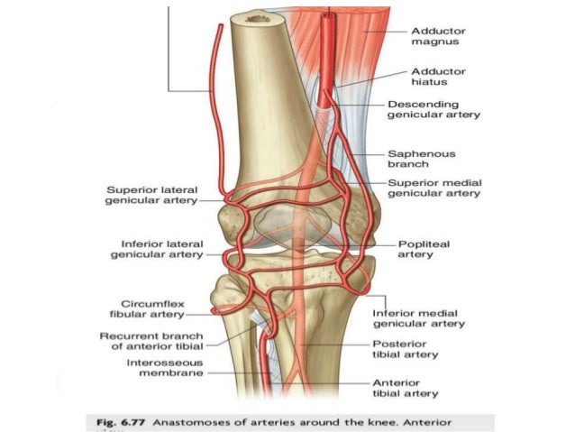 Anatomy And Clinical Importance Of Knee Joint
 Posterior Knee Anatomy Illustrations Image Radiopaedia Org
The Anatomy Of The Posterior Aspect Of The Knee An Anatomic
 Posterior View Of The Right Knee
 Pcl Tear Brisbane Knee And Shoulder Clinic Dr
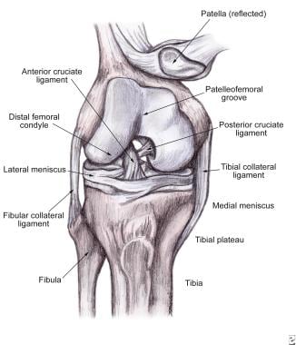 Soft Tissue Knee Injury Practice Essentials Background
 Torn Meniscus Picture Image On Medicinenet Com
 Posterior Knee Anatomy Gray S Anatomy Illustration
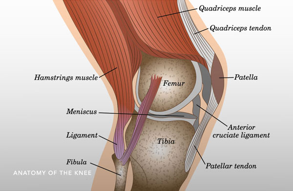 Posterior Cruciate Ligament Pcl Tear Reconstruction
 Knee Anatomy Including Ligaments Cartilage And Meniscus
 Anterior And Posterior Aspects Of The Knee Netter
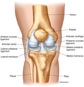 Skeletal System Human Anatomy Life Science Biomedical
 Muscles Of The Posterior Leg Attachments Actions
 Acute Knee Injuries Use Of Decision Rules For Selective


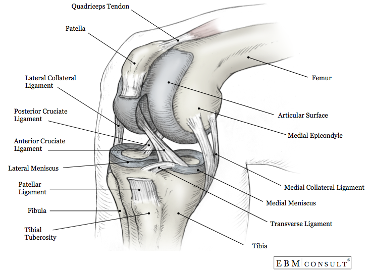

Belum ada Komentar untuk "Posterior Knee Anatomy"
Posting Komentar