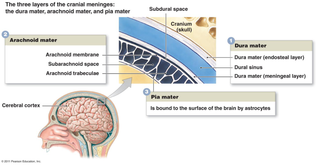
Dural Anatomy
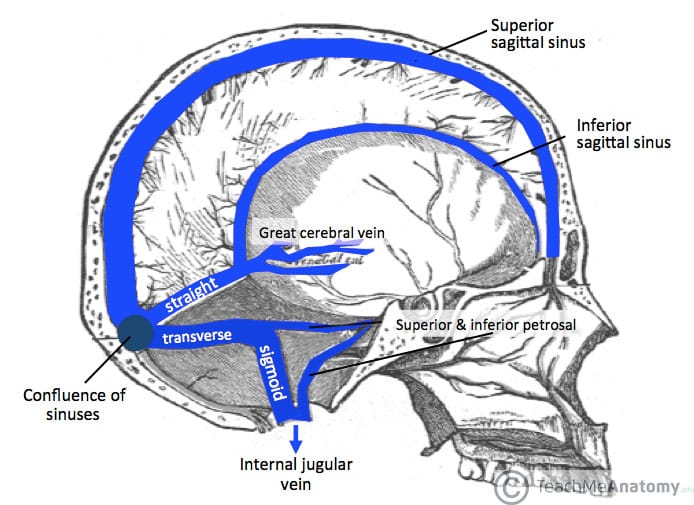
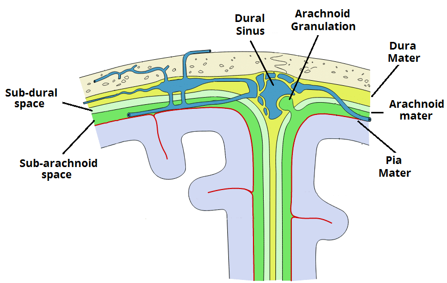
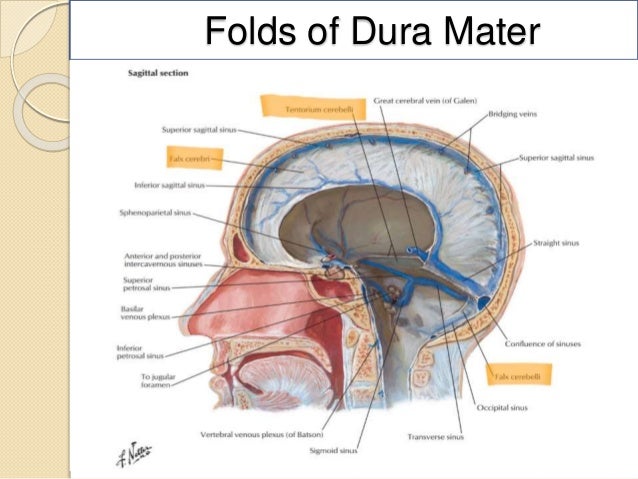
They receive blood from the cerebral veins receive cerebrospinal fluid csf from the subarachnoid space via arachnoid granulations and mainly empty into the internal jugular vein. The tentorium cerebelli exists between and separates the cerebellum and brainstem from the occipital lobes of the cerebrum.
 Meninges And Dural Folds Diagram Quizlet
Following a prologue to dural venous sinuses this article focuses on arachnoid granulations tributaries and the drainage pattern of the venous sinuses.

Dural anatomy. They receive blood from internal and external veins of the brain receive cerebrospinal fluid csf from the subarachnoid space via arachnoid granulations and mainly empty into the internal jugular vein. There are two main dural reflections. The dural venous sinuses also called dural sinuses cerebral sinuses or cranial sinuses are venouschannels found between the endosteal and meningeal layers of dura mater in the brain.
The dural venous sinuses also called dural sinuses cerebral sinuses or cranial sinuses are venous channels found between the endosteal and meningeal layers of dura mater in the brain. There are two sagittal sinuses that occupy the longitudinal cerebral fissure. The sphenoparietal sinus courses along the free border.
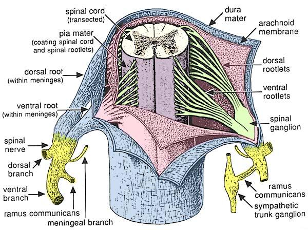
Each anterior cerebral vein leaves the longitudinal cerebral fissure inferiorly. The falx cerebri which separates the two hemispheres of the brain is located in the longitudinal cerebral fissure between the. The dural sac and closely adherent arachnoid mater on its inner surface are a barrier to drug migration into and out of the cerebrospinal fluid during epidural anesthesia.
The termination of the sac well below the conus medullaris classically found at l1 in the adult also provides a degree of safety when performing spinal anesthesia. The dural sac contains the anterior and posterior spinal nerve roots collectively know as the cauda equina. Spinal nerves as there are eight cervical spinal nerves and seven vertebrae the nerves in this region only are numbered according to the vertebra below.
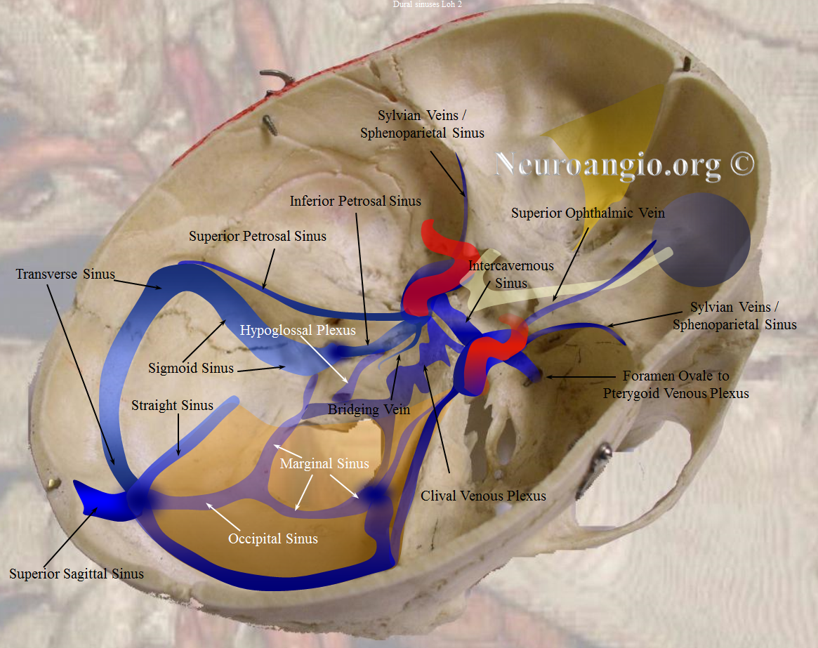
Dural venous sinuses sagittal sinuses. The absence of lymphatic drainage in the brain places the venous outflow system on a pedestal of prime importance. Anatomy and function of the dural venous sinuses.
Pediagenosis
 Ch 12 Gross Anatomy Of The Spinal Cord
 Anatomy Of The Nervous System Microbiology
 Anatomy Imaging And Surgery Of The Intracranial Dural
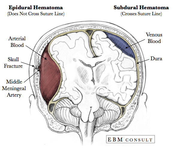 Anatomy Epidural Vs Subdural Hematoma Image
 Dural Venous Sinuses Wikipedia
 Scalp Skull And Meninges The Big Picture Gross Anatomy
 Dural Venous Sinuses
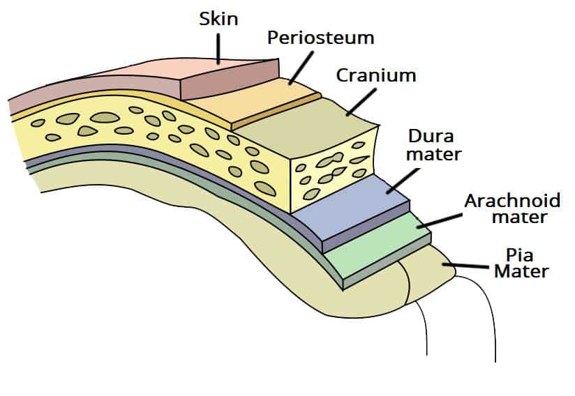 The Meninges Dura Arachnoid Pia Teachmeanatomy
 Dura Mater Radiology Reference Article Radiopaedia Org
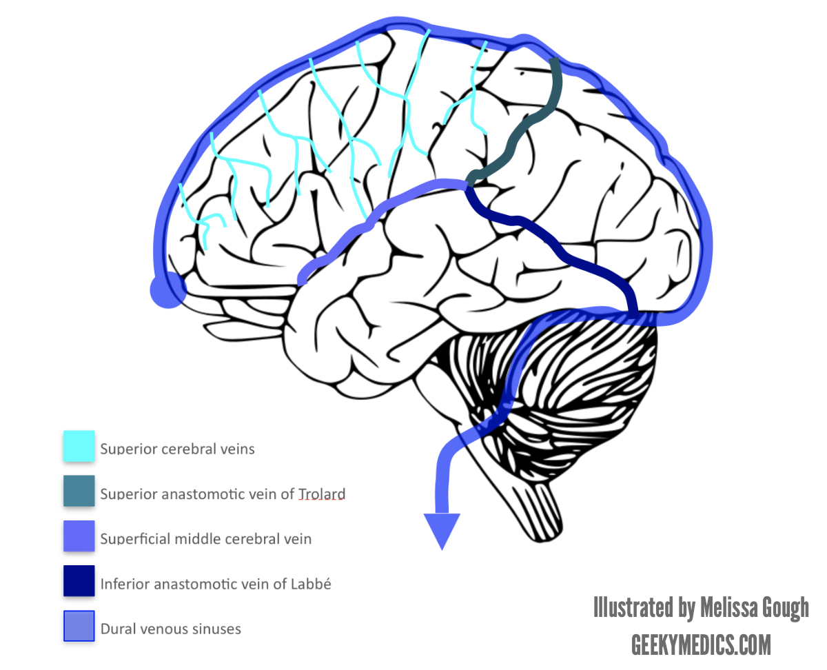 Venous Drainage Of The Brain Anatomy Geeky Medics
 Neuroanatomy Online Lab 4 External And Internal Anatomy
 Venous Drainage Of The Cns Cerebrum Teachmeanatomy
 Dural Venous Sinuses 3d Anatomy Tutorial
 File Three Main Variations Of The Venous Anatomy Of The

 Segregation Of Dural Vessel By Fit Parameters And Anatomy A
 Spinal Cord Anatomy Spine Orthobullets
 Meninges Dura Mater Arachnoid Mater Pia Mater
 Figure 1 From Surgical Anatomy Of The Dural Walls Of The
 Ch 12 Cerebrospinal Fluid
![]() Meninges Of The Brain And Spinal Cord Anatomy Function
Meninges Of The Brain And Spinal Cord Anatomy Function

![]() The Meninges
The Meninges
 Structure And Functions Of The Dura Mater Explained With
 Dural Venous Sinuses
 The Meninges Dura Arachnoid Pia Teachmeanatomy
 Amazon Com Anatomy Dural Venous Spine Print Sra3 12x18
 Meninges An Overview Sciencedirect Topics
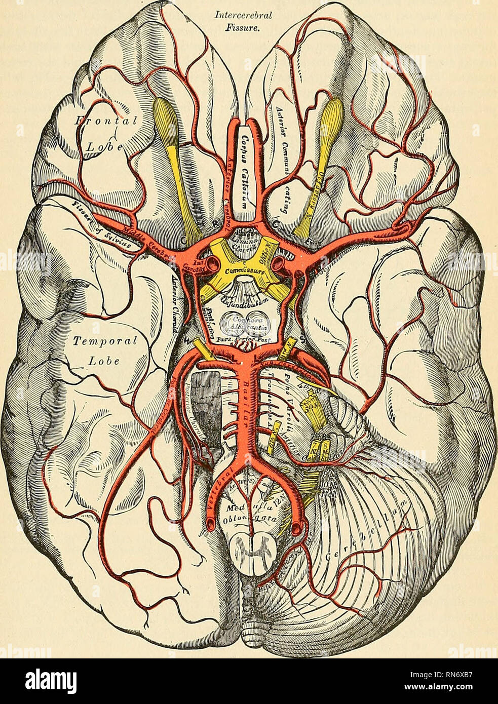 Anatomy Descriptive And Applied Anatomy The Internal
 Meninges Dura Mater Pia Mater Arachnoid Mater Brain Brain
 Cranial Cavity Department Of Anatomy
Belum ada Komentar untuk "Dural Anatomy"
Posting Komentar