Knee Tendon Anatomy
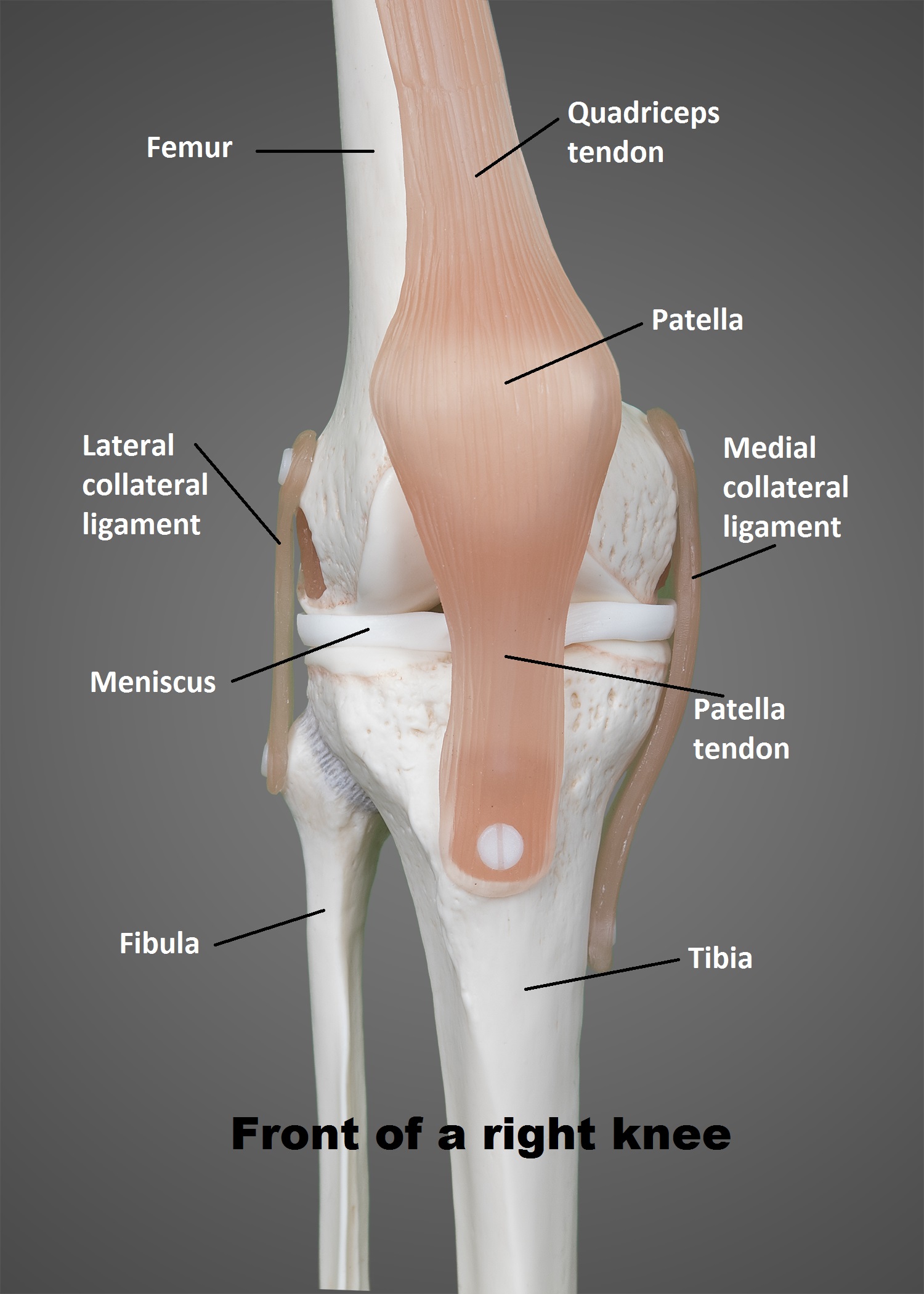
The knee is a hinge joint that is responsible for weight bearing and movement. Femur the upper leg bone or thigh bone.
 Knee Anatomy
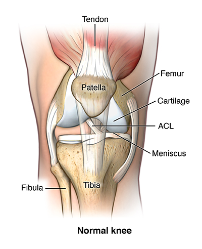
Articular cartilage allows the knee bones to move easily as the knee bends and straightens.

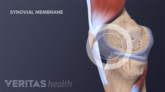
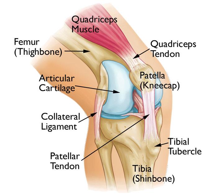
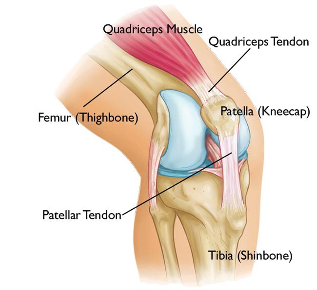
Knee tendon anatomy. There is articular cartilage anywhere that two bony surfaces come into contact with each other. In the knee articular cartilage covers the ends of the femur the femoral groove the top of the tibia and the underside of the patella. There are two major tendons in the kneethe quadriceps and patellar.
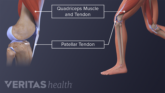
Ligaments are tough fibrous connective tissues which link bone to bone made of collagen. The largest joint in the body the knee moves like a hinge allowing you to sit squat walk or jump. The quadriceps tendon connects the quadriceps muscles of the thigh to the kneecap and provides the power for straightening the knee.
The two important tendons in the knee are 1 the quadriceps tendon connecting the quadriceps muscle which lies on the front of the thigh to the patella. When there is damage to one of the structures that surrounds the knee joint this can lead to discomfort and disability. The knee consists of three bones.
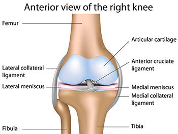
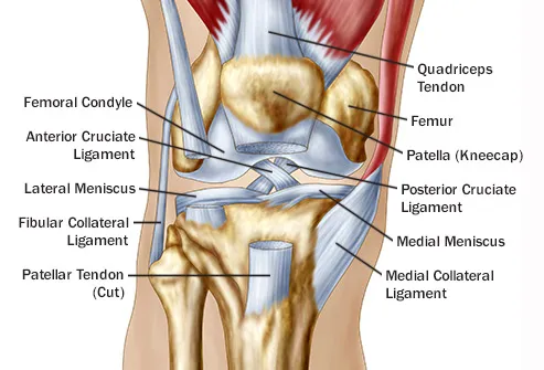
In knee joint anatomy they are the main stabilising structures of the knee acl pcl mcl and lcl preventing excessive movements and instability. It also helps hold the patella in the patellofemoral groove in the femur. The smaller bone that runs alongside the tibia fibula and the kneecap patella are the other bones that make the knee joint.
Knee anatomy share on pinterest the knee is the most complex joint in the human body. The knee joint is a complex structure that involves bones tendons ligaments muscles and other structures for normal function. Tibia the bone at the front of the lower leg or shin bone.
The knee is one of the largest and most complex joints in the body. Answer tendons connect muscles to bones. Tendons in the knee.
The most common ligament injuries are acl tears mcl tears. Tendons connect the knee bones to the leg muscles that move the knee joint. The knee is the joint where the bones of the lower and upper legs meet.
The knee joins the thigh bone femur to the shin bone tibia.
Anatomy Causes And Treatment Of Jumper S Knee Patellar
 Achilles Tendon Human Anatomy Picture Definition

 Knee Joint Anatomy Motion Knee Pain Explained
 Tendon Stem Cells Could Revolutionize Injury Recovery
 Patellar Tendinitis Symptoms And Causes Mayo Clinic
 Leg Knee Anatomy
 Knee Joint Anatomy Bones Ligaments Muscles Tendons Function
 Patellofemoral Pain Syndrome Orthoinfo Aaos
Common Knee Injuries Orthoinfo Aaos
 Understanding The Anatomy Of The Knee Bodyheal
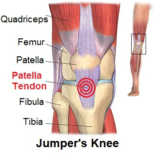 Patellar Tendonitis Jumpers Knee Symptoms Diagnosis
/188058334-crop-56aae7425f9b58b7d0091480.jpg) What Is Causing Your Knee Pain
What Is Causing Your Knee Pain
 Quadriceps Tendonitis Of The Knee Richmond Va Sports Medicine
 Understanding Jumper S Knee
 Knee Wikipedia

 The Knee Patella Tendinopathy
 Reasons For Pain Behind In Back Of The Knee

Common Knee Injuries Orthoinfo Aaos
 Medial Patellofemoral Ligament Mpfl Reconstruction
 A Guide To Your Knees Well Guides The New York Times
 Knee Injuries And Disorders Richmond Va Knee Anatomy
 The Knee Ut Health San Antonio
 Knee Ligaments Joi Jacksonville Orthopaedic Institute
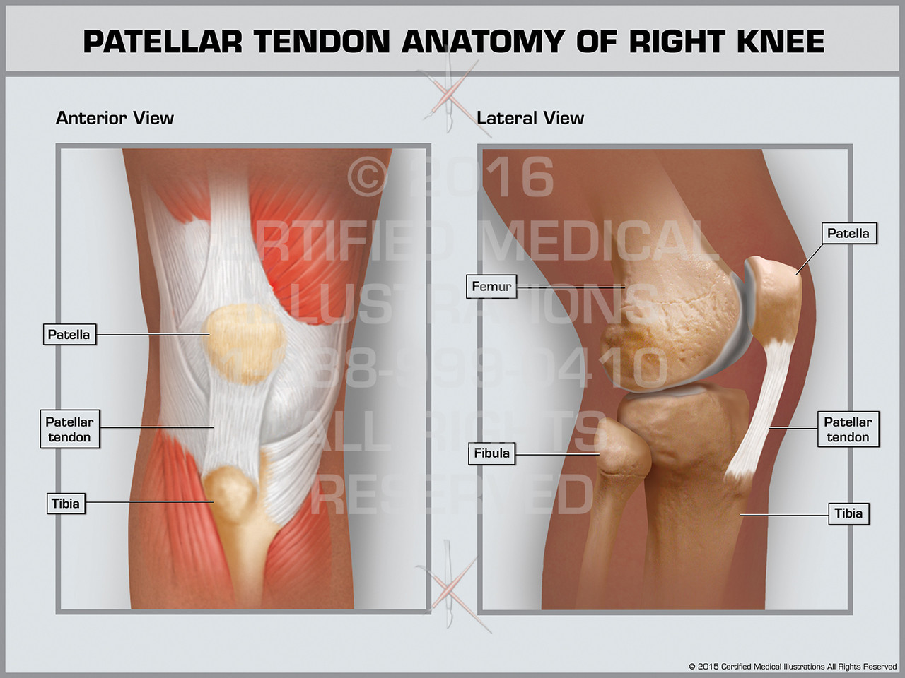 Patellar Tendon Anatomy Of Right Knee
 Knees Ligament And Tendon Pains Bing Images Knee
 Adolescent Anterior Knee Pain Orthoinfo Aaos
Belum ada Komentar untuk "Knee Tendon Anatomy"
Posting Komentar