Thoracic Duct Anatomy
It is also called the left lymphatic duct or the alimentary duct. Multiple ducts 20 1 2 4.
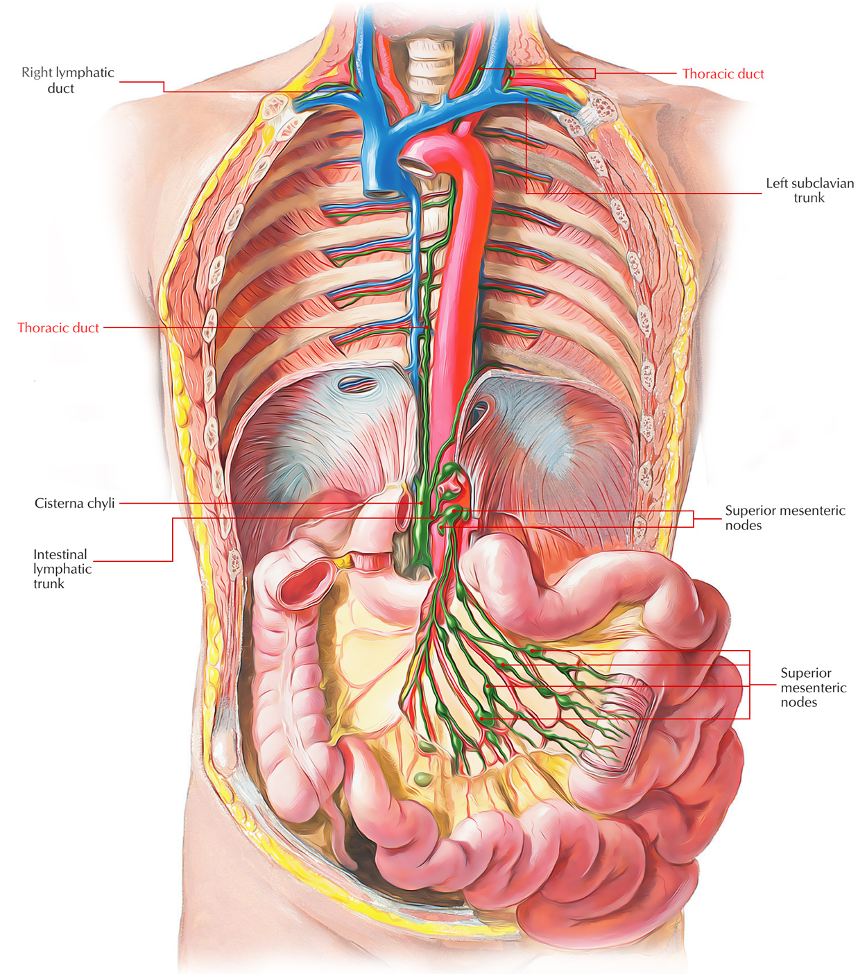 Easy Notes On Thoracic Duct Learn In Just 4 Minutes
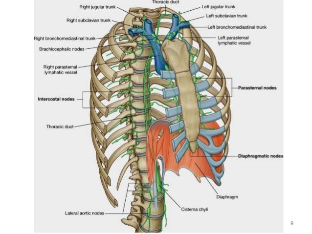
The thoracic duct is the common channel draining lymphatic fluid from the body to the venous system with the exception of the right face and neck right arm right thorax and lung and convex surface of the liver.

Thoracic duct anatomy. Thoracic duct in mammalian anatomy a principal channel for lymph. The thoracic duct carries chyle a liquid containing both lymph and emulsified fats rather than pure lymph. From about the level of the small of the back it runs up through the body close in front of the backbone to the base of the neck where it opens into a blood vessel at the point at which the left subclavian vein and the left internal jugular vein join to form the left brachiocephalic vein.
The thoracic duct is a tubular structure that is 2 3 mm in diameter. The thoracic duct extends from the twelfth thoracic vertebra to the root of the neck. It length varies between 38 45 cm and it is 2 3 mm in diameter.
Thoracic duct anatomy must be understood in the context of its embryology. The thoracic duct begins in the abdomen by a triangular dilatation the cisterna chyli in front of l2 vertebral body. The thoracic duct also known as van hoornes canal is the largest lymphatic vessel of the lymphatic system of the body.
A large portion of the bodys lymph is collected by this duct and then drained into the bloodstream near the brachiocephalic vein between. Thoracic duct anatomy overview. Laceration of the thoracic duct during lung surgery results in chyle entering into the pleural cavity producing a clinical condition named chylothorax.
The thoracic duct has variant anatomy in 40 range 30 50 of the population 1 4. The structure of the thoracic duct is considered to be similar to that. Gross anatomy of thoracic duct in abdomen.
The thoracic duct is the largest lymphatic vessel within the human body and plays a key role in the lymphatic system. It is approximately 40 cm in length in adults and approximately 5 mm in width at its abdominal origin. Double thoracic ducts ie.
In human anatomy the thoracic duct is the larger of the two lymph ducts of the lymphatic system. The other duct is the right lymphatic duct. It is also known as the left lymphatic duct alimentary duct chyliferous duct and van hoornes canal.
Left internal jugular vein left external jugular vein azygos vein brachiocephalic vein or left subclavian vein 1 4. Anatomy of the thoracic duct the usual course of the thoracic duct is illustrated in figure 66 1. The thoracic duct is a tubular structure that extends from l2 vertebra to the root of the neck.
The thoracic duct is thin walled and might be colorless for that reason its sometimes injured inadvertently during surgical procedures in the posterior mediastinum.
 Thoracic Duct Anatomical Vector Illustration Diagram
 Thoracic Duct An Overview Sciencedirect Topics
 Thoracic Duct Anatomy Course Relations Tributaries And Clinical Significance Usmle Step 1
 Thoracic Duct Anatomical Vector Illustration Diagram Medical Scheme Canvas Print
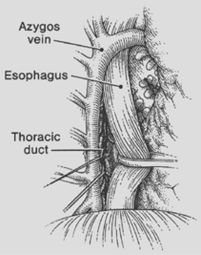 Anatomy Of The Thoracic Duct And Chylothorax Thoracic Key
 Naturaltherapy Lymphatic Drainage Ginger Oil Lymphatic
 Thoracic Duct Anatomical Vector Illustration Diagram Stock
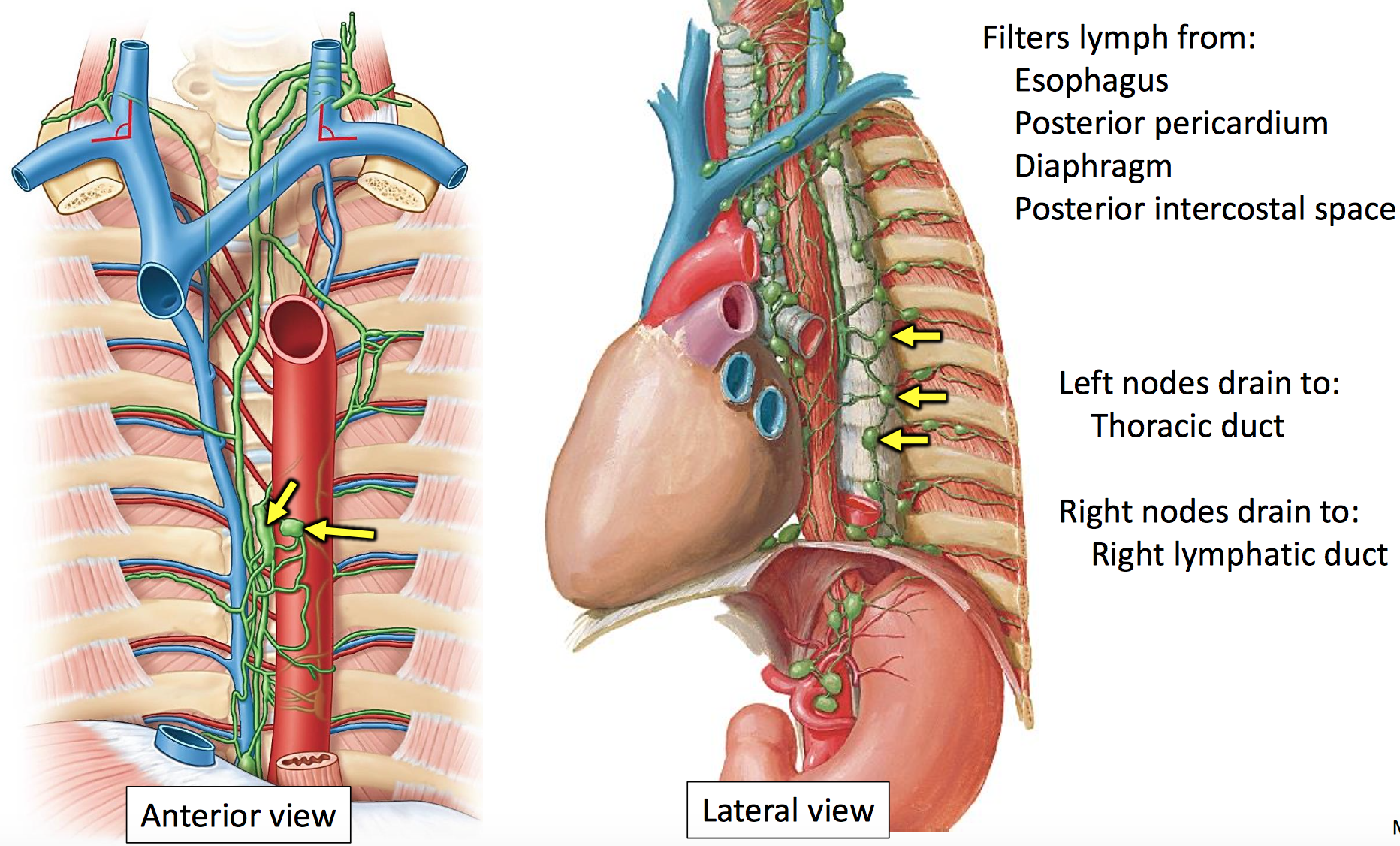
 Anterior Thoracic Wall The Big Picture Gross Anatomy 2e
:watermark(/images/watermark_only.png,0,0,0):watermark(/images/logo_url.png,-10,-10,0):format(jpeg)/images/anatomy_term/ductus-thoracicus-2/ypokyGBKCr1ggLyTmb8yuQ_Ductus_thoracicus_1.png) Thoracic Duct Anatomy Course And Clinical Significance
Thoracic Duct Anatomy Course And Clinical Significance
 Latest 1402 953 Thoracic Duct Lymphatic System
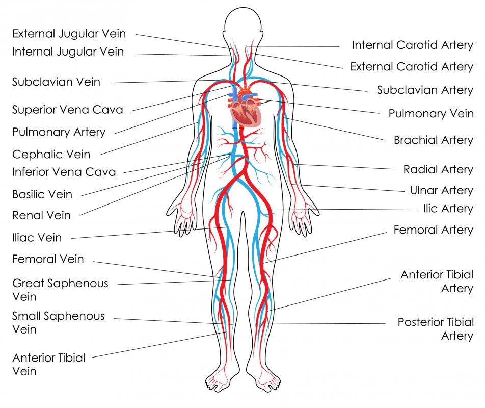 What Is The Thoracic Duct With Pictures
 Section 5 Lymphatic System Diagram Quizlet
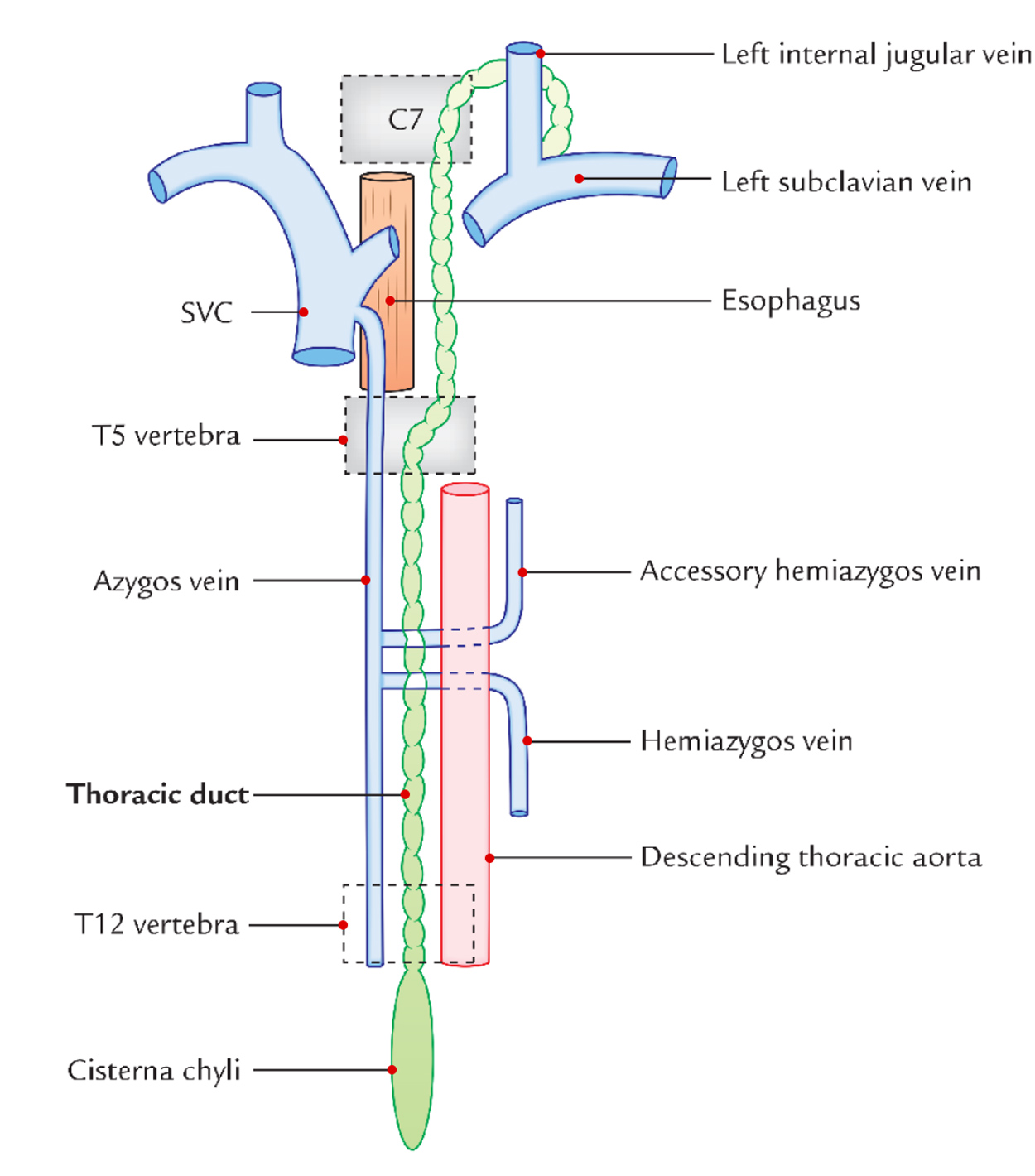 Easy Notes On Thoracic Duct Learn In Just 4 Minutes
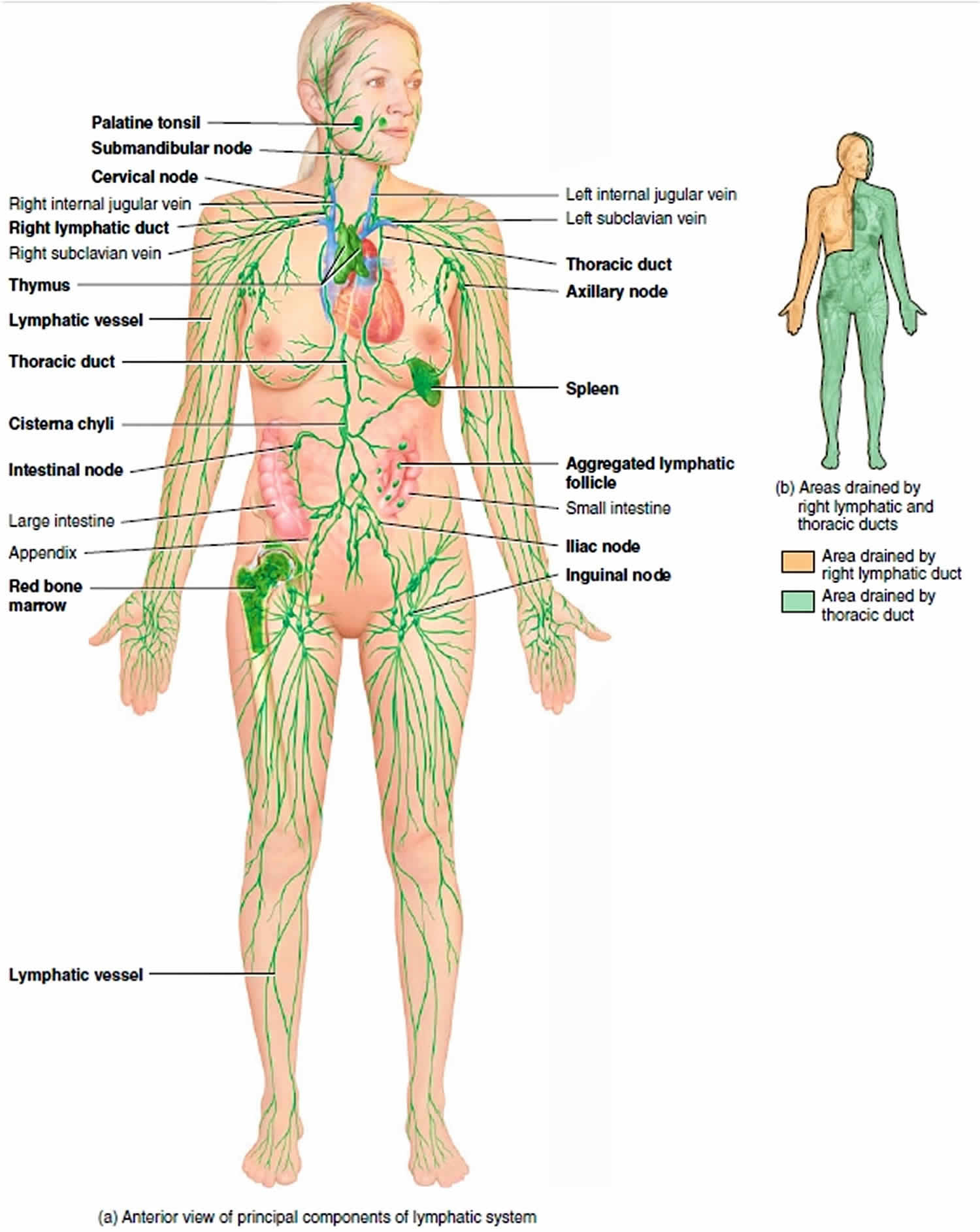 Thoracic Duct Anatomy Thoracic Duct Drainage Function
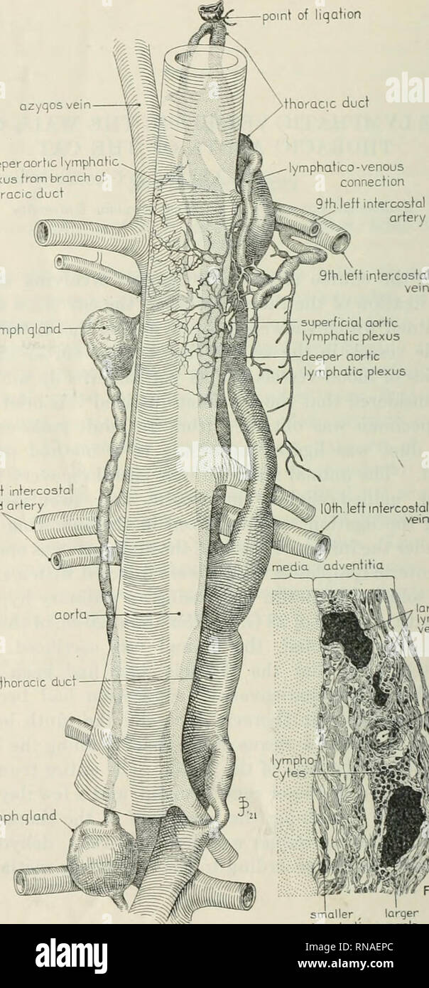 The Anatomical Record 1922 1923 Anatomy 344 Qzyqos Vein
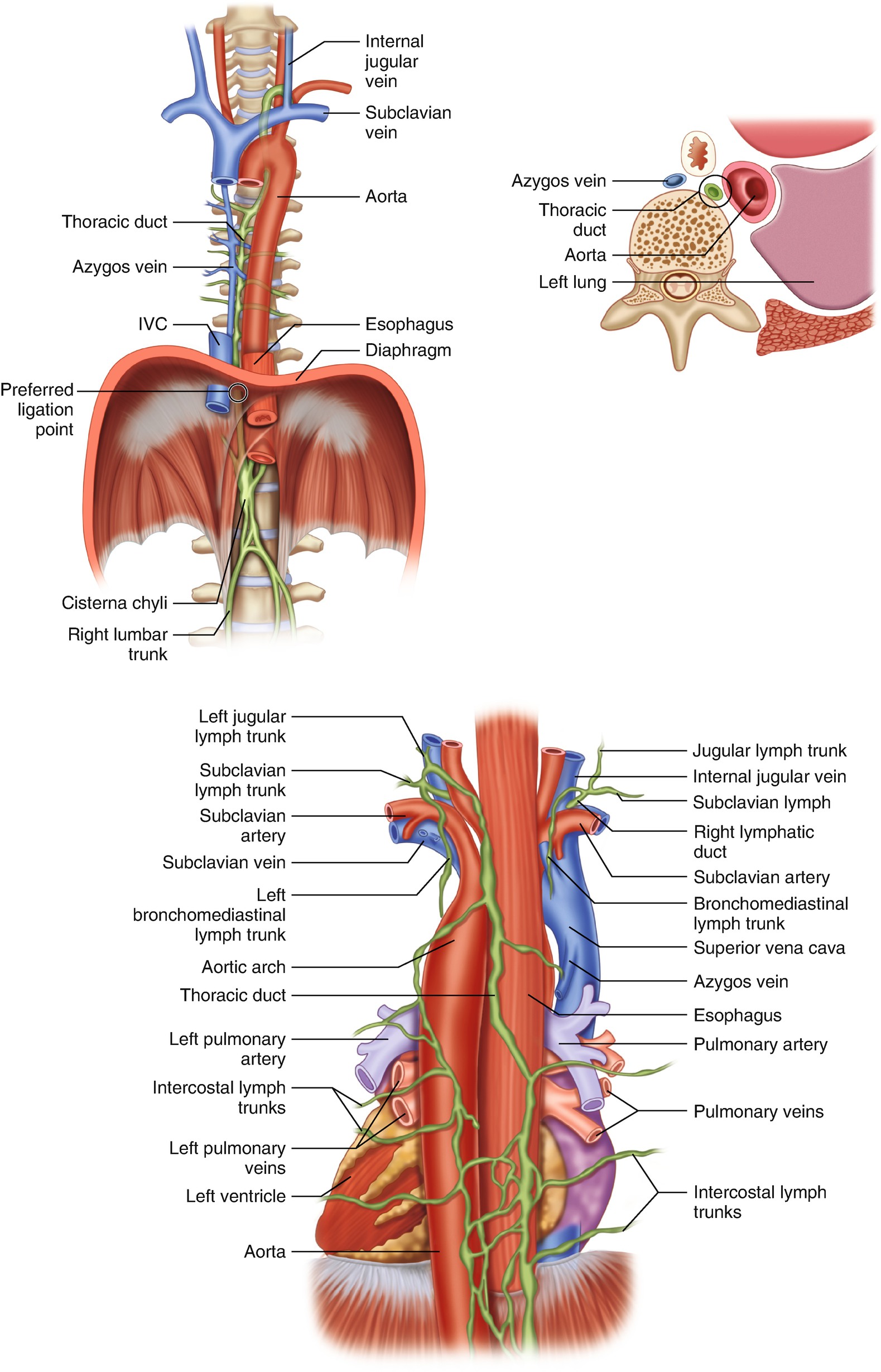 Robotic Transthoracic Thoracic Duct Ligation Springerlink
 Review Of Thoracic Duct Anatomical Variations And Clinical
 Pin By Valerie On Anatomy And Physiology Thoracic Duct
:watermark(/images/logo_url.png,-10,-10,0):format(jpeg)/images/anatomy_term/vena-subclavia-sinistra/jqk9icwH8BbntPN05YTQGg_V._subclavia_sinistra_1.png) Thoracic Duct Anatomy Course And Clinical Significance
Thoracic Duct Anatomy Course And Clinical Significance




Belum ada Komentar untuk "Thoracic Duct Anatomy"
Posting Komentar