Brain And Spinal Cord Anatomy
The senses taste smell sight hearing touch emotions thoughts and movement are controlled by the brain. This article discusses the spinal cords anatomy and potential signs and symptoms that can develop if cord compression or injury occurs at the level of the cervical spine.
The spinal cord is critical for transmitting information to and from the brain.
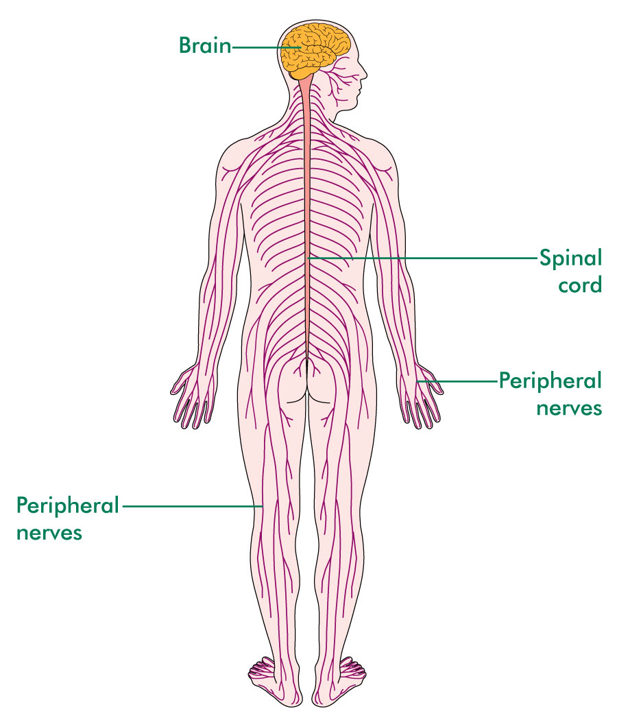
Brain and spinal cord anatomy. Learn vocabulary terms and more with flashcards games and other study tools. The spinal cord is located immediately below the brain stem. Gross anatomy the spinal cord is part of the central nervous system cns which extends caudally and is protected by the bony structures of the vertebral column.
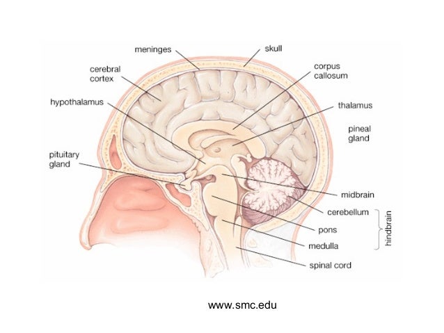
Spinal cord anatomy basically spinal cord is a long and narrow bundle of nervous tissues and support cells which extends from the base of our brain to the upper lumbar region. This allows the brain to send messages throughout the body. Anatomy of the brain and spinal cord the brain is an organ located in the skull.
The central nervous system cns is composed of the brain and spinal cord. It extends through the foramen magnum a hole at the base of the skull. Welcome to headneckbrainspine a website intended for those interested in neuroradiology anatomy and learning from neuroradiology cases.
Start studying anatomy brain and spinal cord. The spinal cord is located immediately below the brain stem. It passes through the spinal canal or spinal cavity of the vertebral column ie the backbone or spine.
Brain and spine tumor anatomy and functions the brain controls many important body functions such as emotions vision thought speech and movement. To navigate the website click on the images below or on the above menu. It is covered by the three membranes of the cns ie the dura mater arachnoid and the innermost pia mater.
It weighs about 3 pounds. Thank you to dr kelly wallace for contributing the knowledge and the demo for the video. Brain and spinal cord review video with terms.
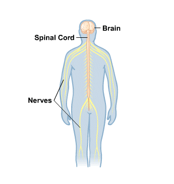
Anatomy of the sheep brain video for anatomy class. The peripheral nervous system pns is composed of spinal nerves that branch from the spinal cord and cranial nerves that branch from the brain. The brain and spinal cord together make up the central nervous system.
The spinal cord connects the brain to nerves in most parts of the body.
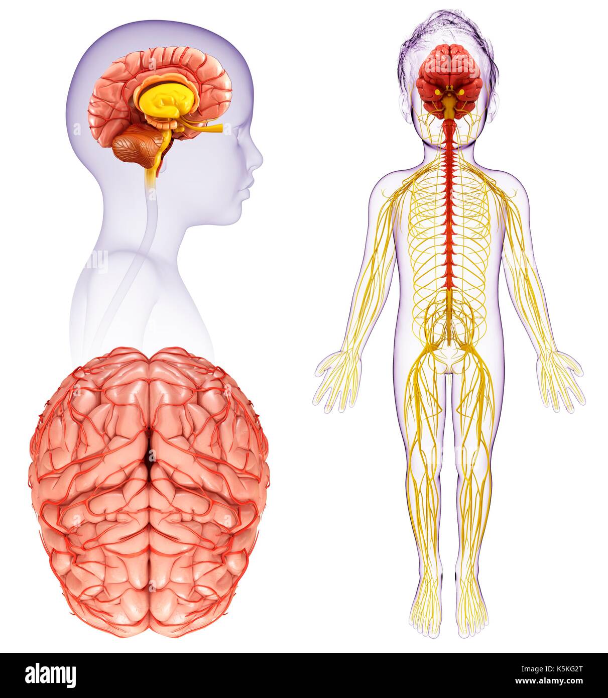 Illustration Of A Child S Brain And Spinal Cord Anatomy
Anatomy Of The Brain The Basics Understanding Brain Injury
 Sci Info Bryon Riesch Paralysis Foundation
 Anatomy Of The Brain And Spinal Cord Seattle Cancer Care
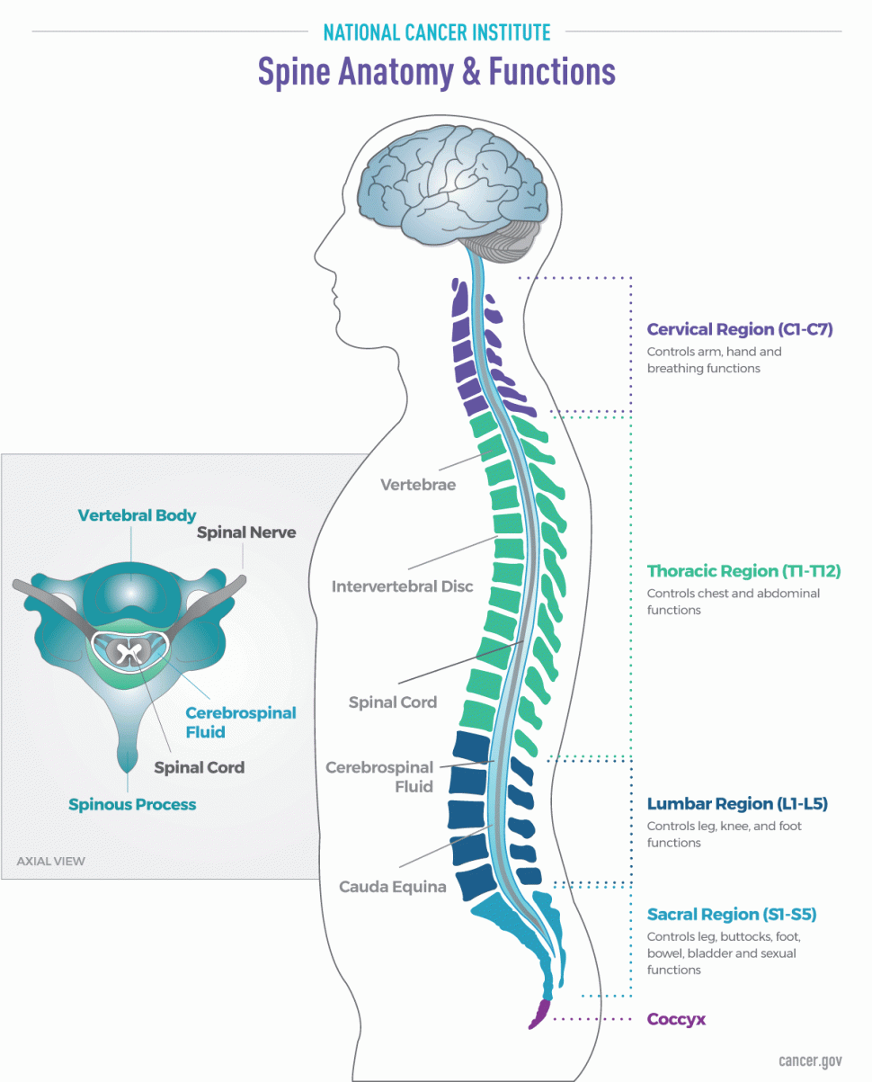 Brain And Spine Tumor Anatomy And Functions The Spine
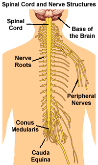 Understanding Spinal Anatomy Spinal Cord And Nerve Roots
 If You Re An Adult With A Brain Or Spinal Cord Tumor
 Brain And Spinal Cord Anatomy Cross Section Stock Photo
What Is The Difference Between Meninges Of Brain And Spinal
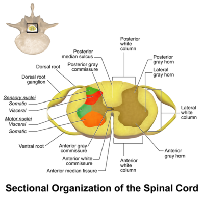 Spinal Cord Anatomy Physiopedia
 Becoming Mindful Of The Brain And Its Functions Igea Brain
 Sagittal Section Of The Brain And Spinal Cord Science
 Labeled Pictures Of The Brain Human Spinal Cord Diagram
 Brain Anatomy Goodman Campbell Brain And Spine
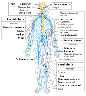 Central Nervous System Disease Wikipedia
 Nervous System Infections Dr Jeannine Durdik
 The Brain And Spinal Cord Learning How We Think Feel And
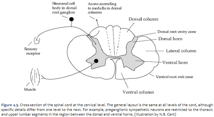 Duke Neurosciences Lab 2 Spinal Cord Brainstem Surface
 Atlas Of The Human Brain And Spinal Cord 9780763753184
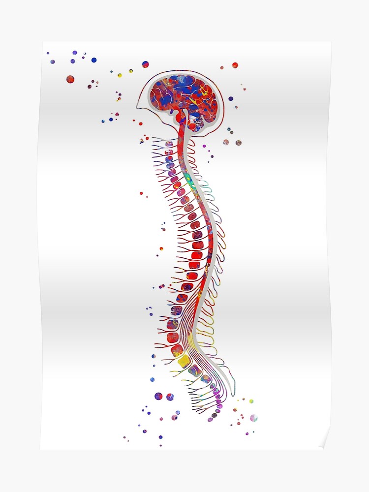 Brain With Spinal Cord Brain Anatomy Medical Art Poster
![]() Brain 101 An Overview Of The Anatomy And Physiology Of The
Brain 101 An Overview Of The Anatomy And Physiology Of The
 Human Anatomy Art Print Brain Spinal Cord Vintage Anatomy Doctor Medical Art Antique Book Plate M Art Print By Frenchfineart
 Spinal Cord Tumours Macmillan Cancer Support
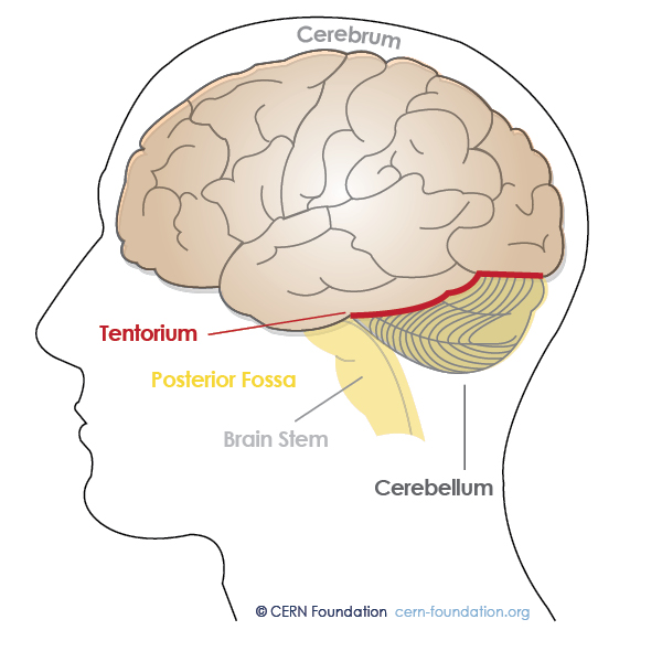 Spine And Brain Tumor And Anatomy Cern Foundation
 Brain Spinal Cord Atlas Of Anatomy

Belum ada Komentar untuk "Brain And Spinal Cord Anatomy"
Posting Komentar