Horse Hock Anatomy
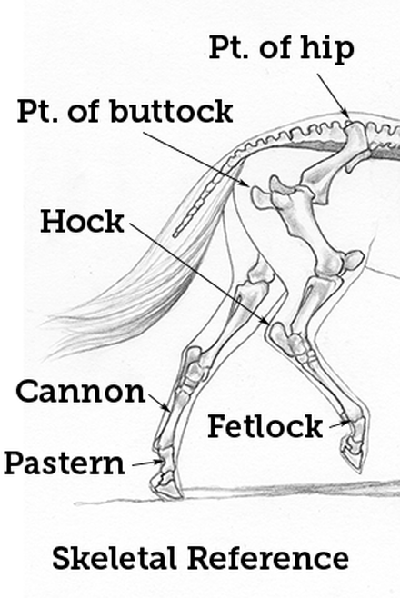
The hock is a horsemans term for the tarsus an anatomic region of the horses hind limb. Equine rear leg bones and function the horse leg anatomy in the rear includes the bones of the pelvis the ilium ischium and pubic bones femur tibia fibula metatarsus and the phalanxes.
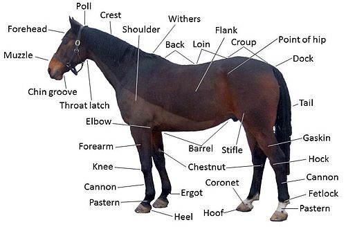 Equine Anatomy Wikipedia
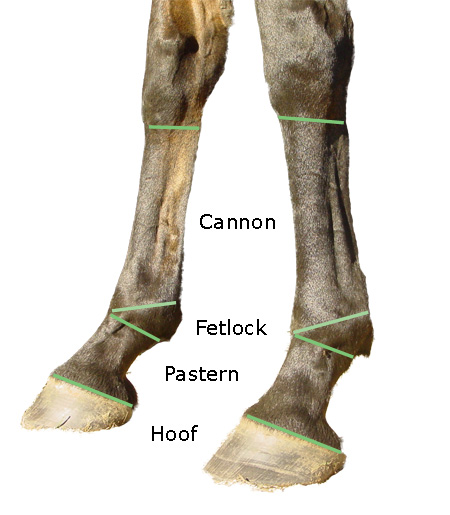
Below the hock joint are the hind cannon with splint bones the long and short pastern the coffin joint and bone the sesamoid bones.

Horse hock anatomy. The horses hock is made up of 10 bones and 4 joints supported by several ligaments. Distal intertarsal joint or centrodistal joint. 1st 2nd fused tarsal b.
There are various disease processes that affect the nature of the synovial fluid because of inflammation and disease in the synovial membrane. The horses hock joint is one of the hardest working of all the joints and plays a critical role especially in performance horses. Tarsal anatomy of the horse 1.
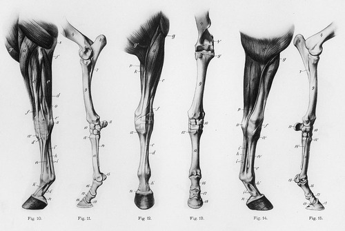
Equine anatomy refers to the gross and microscopic anatomy of horses and other equids including donkeys and zebras. While all anatomical features of equids are described in the same terms as for other animals by the international committee on veterinary gross anatomical nomenclature in the book nomina anatomica veterinaria there are many horse specific colloquial terms used by equestrians. It also includes the joints of the hip stifle hock fetlock pastern and coffin.
Tibiotarsal or tarsocrural joint. Tarsus anatomy dane tatarniuk dvm 2. The horses hind limbs.
It also includes the joints of the hip stifle hock fetlock pastern and coffin. Inflammation in the joint causes excessive fluid production. In the horse the hock consists of multiple joints namely.
The synovial membrane secretes the synovial fluid which provides lubrication within the joint. The horse leg anatomy in the rear includes the bones of the pelvis the ilium ischium and pubic bones femur tibia fibula metatarsus and the phalanxes. Proximal intertarsal joint or talocalcanealcentroquartal joint.
Bones talus b. The horses hock joint horses hock anatomy. 2nd 3rd 4th metatarsal b.
It is also one of the most complicated. Horses of all breeds types and disciplines can suffer from hock related lameness problems especially those that work heavily off of their hind limbs. The hock joint allows movement of the hind leg and consists of the tarsus bones the tuber and the calcaneus at the back which forms the point of the hock.
 Tarsus Aovet Equine Ao Surgery Reference
 The Horse S Hock Treatments And Symptoms Of Hock Joint
Equine Hock Anterior View Horse Anatomy Horse Bones
 Horse Anatomy Mobility Health
 Horse Hock Anatomy Horse Anatomy Animal Medicine Horse Care
 Equine Anatomy Hock Veterinary Radiology Horse Care
 Equine Distal Limb Diagnostic Anaesthesia 1 Basic
 Horse Anatomy Hock Injury Veterinarian Chart 1920s Color Lithograph Antique Large Animal
 Horse Leg Anatomy Learn Everything You Did Not Know Medrego

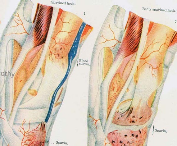 Horse Anatomy Hock Injury Veterinarian Chart 1920s Color Lithograph Antique Large Animal
 Regional Anesthesia In Equine Lameness Musculoskeletal
 Horse Bones Everything You Need To Know To Get Started

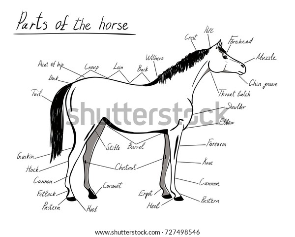 Parts Horse Equine Anatomy White Black Stock Vector Royalty
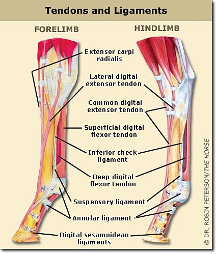
 Tendons And Ligaments Structure And Injury Rainland Farm

 Importance Of Proper Hind Leg Conformation Equimed Horse
 Reading Radiographs Horse Rider
 Sesamoid Injuries In Horses Diagnosis Treatment And
 Horse Anatomy Mobility Health
 5 Common Sport Horse Injuries Expert How To For English Riders
 Causes Of Equine Lameness Equimed Horse Health Matters
 Luxation Of The Superficial Digital Flexor Tendon From The
 Novobrace Tendonitis Desmitis And Soft Tissue Injury
Belum ada Komentar untuk "Horse Hock Anatomy"
Posting Komentar