Mri Anatomy Knee
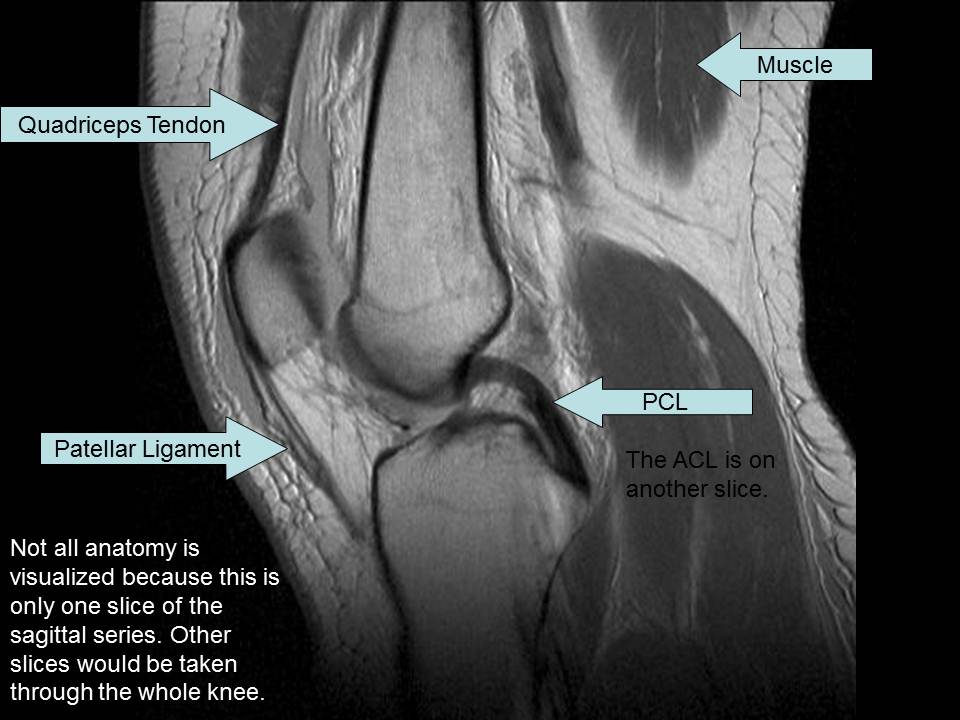
Through the use of magnetic resonance imaging clinicians can diagnose ligament and meniscal injuries along with identifying cartilage defects bone fractures and bruises. Each anatomical structure is labelled interactively.
Knee seems like it should be pretty easy right.

Mri anatomy knee. Colorado knee specialist dr. This atlas of cross sectional anatomy of the knee is based on imagery by magnetic resonance mri. Normal mri anatomy of the knee 639.
Anatomy of the knee mri atlas of the human body using cross sectional imaging. Robert laprade discusses how to read an mri of a normal knee. Use the mouse scroll wheel to move the images up and down alternatively use the tiny arrows on both side of the image to move the images.
After all an entire year of fellowship training is dedicated to musculoskeletal imaging. The iliotibial band bursa is situated between the tibia and distal iliotibial band immedi ately proximal to its insertion on gerdys tubercle. Atlas of knee mri anatomy.
By now you probably know that the anatomy is deceptively complex combinations of injuries can be challenging and of course the referring clinicians expectations are as high as the range of meniscus injuries is wide. This mri knee sagittal cross sectional anatomy tool is absolutely free to use. Anatomy of the knee can be complicated and hard to understand.
The fcl biceps femoris bursa is found lateral to the distal fcl and insinuates anterior and antero medial in relation to this ligament. Use the mouse to scroll. This webpage presents the anatomical structures found on knee mri.
Click on a link to get t1 coronal view t2 fatsat axial view t2 fatsat coronal view t2 fatsat sagittal view. This tool is at the same time useful for the training and teaching of the anatomy. Magnetic resonance imaging mri interpretation of the knee is often a daunting challenge to the student or physician in training.
 The Knee Mri Atlas Of Anatomy In Medical Imagery
 Radiologic Evaluation Of The Knee Fundamentals Of
Stanford Msk Mri Atlas C 2019
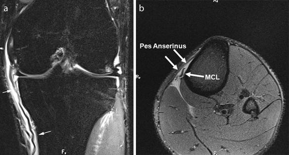
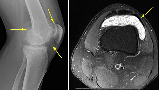
 Fat Pad Impingement Causes And Treatment Uw Health
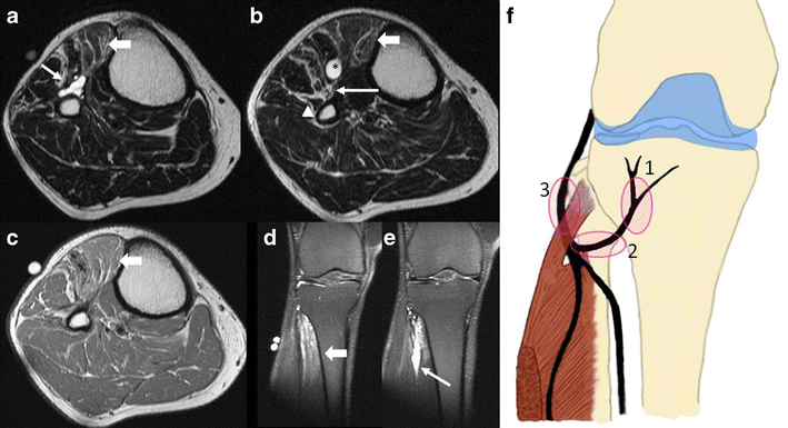
 Mri Of The Knee What Do We Miss Springerlink

 Knee Wikidoc

 Mri Knee Anatomy Knee Sagittal Anatomy Free Cross
 Knee Ligament Anatomy Images Stock Photos Vectors
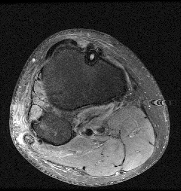
 Example Of Mri Slice Mid Thigh With The Knee Extensors And


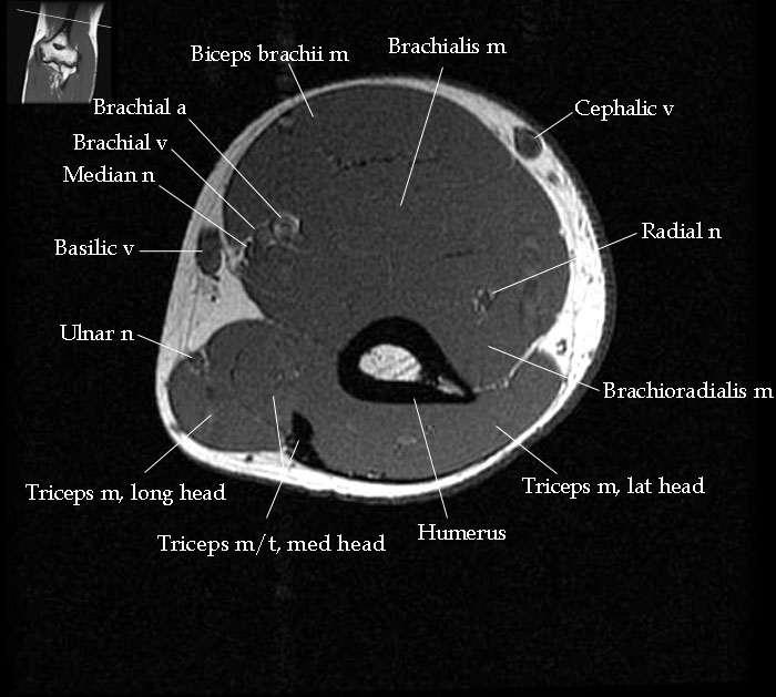 Mri Elbow Anatomy
 Synovial Chondromatosis Orthoinfo Aaos
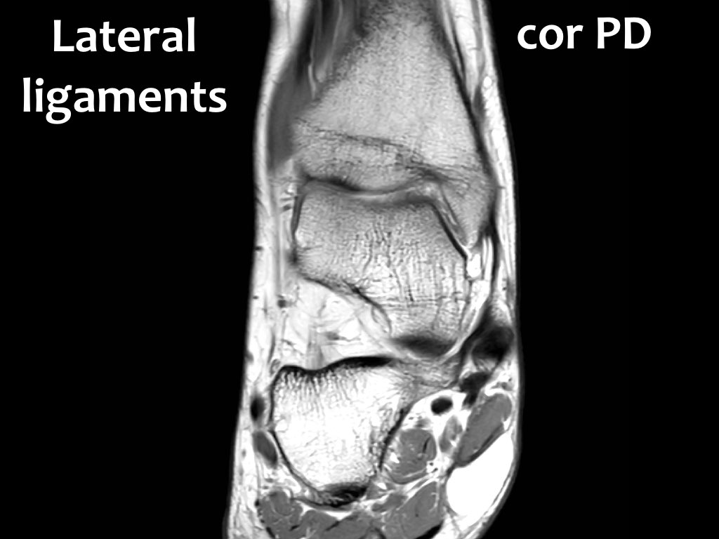 The Radiology Assistant Ankle Mri Examination

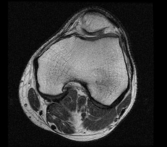
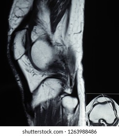
 Anatomy Quiz Mri Knee Radiology Case Radiopaedia Org
 Knee Anatomy Mri Knee Coronal Anatomy Free Cross
 Mri Knee Anatomy Knee Sagittal Anatomy Free Cross
 Peroneal Nerve Normal Anatomy And Pathologic Findings On
 Knee Anatomy Mri Knee Coronal Anatomy Free Cross
 The Knee Mri Atlas Of Anatomy In Medical Imagery

 Mri Knee Joint Anatomy
 Knee Anatomy Mri Knee Coronal Anatomy Free Cross
Mri Knee Anatomy Knee Sagittal Anatomy Free Cross
 Mri Anatomy Of Knee Dr Muhammad Bin Zulfiqar
 Department Of Anatomy Med Univ Of Warsaw Poland Knee
Belum ada Komentar untuk "Mri Anatomy Knee"
Posting Komentar