Sphenoid Sinus Anatomy
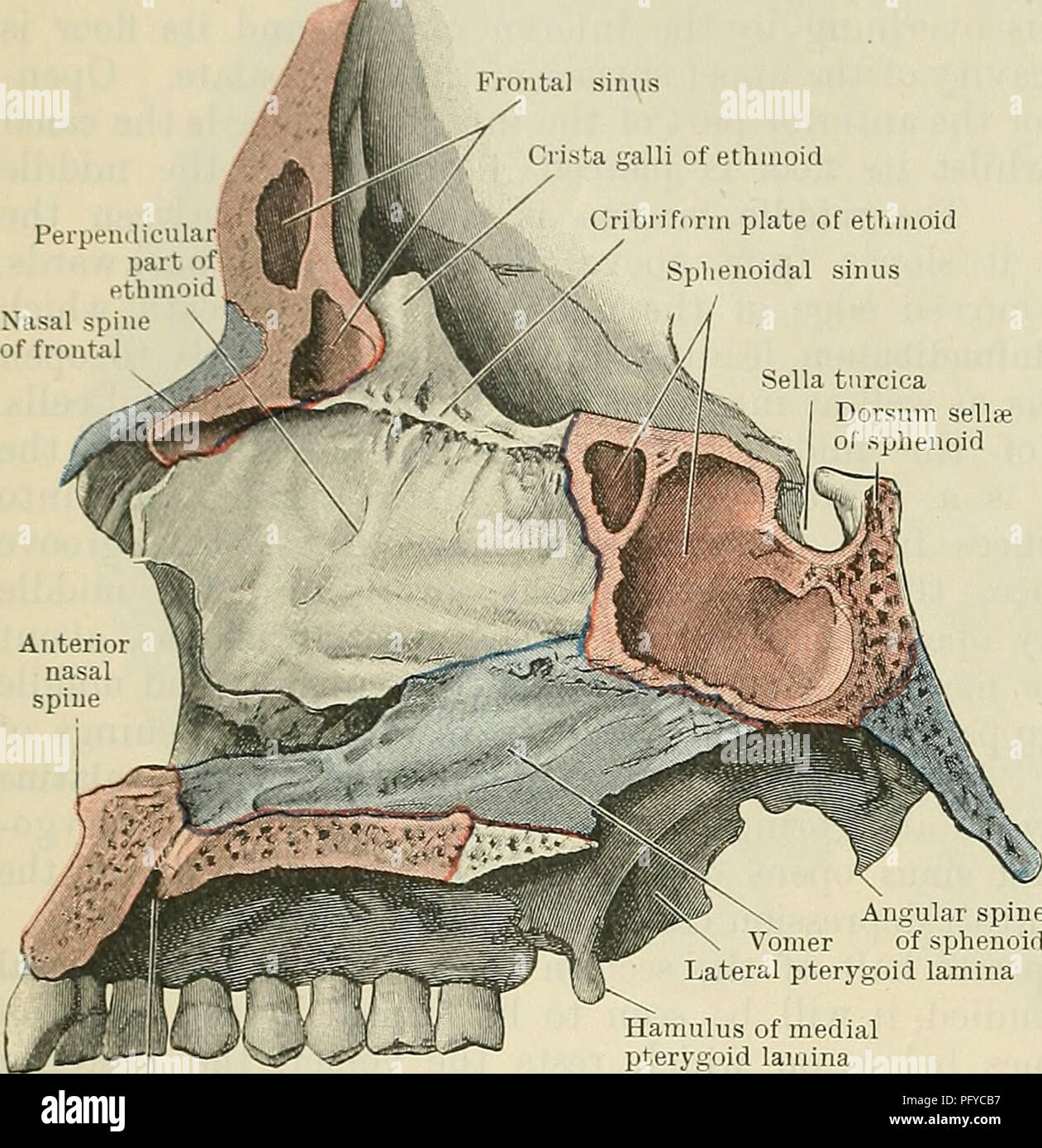
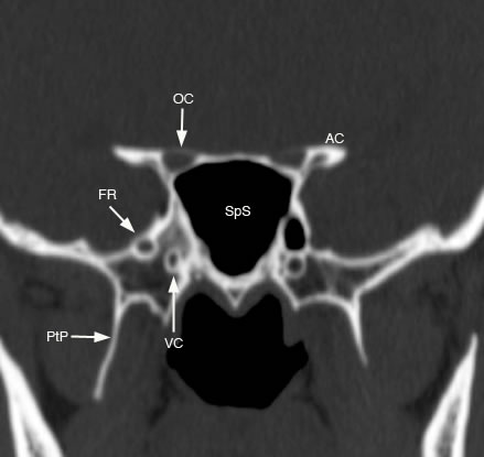
The sphenoid sinuses are paired spaces formed within the body of the sphenoid bone communicating with the roof of the nasal cavity via the sphenoethmoidal recess in its anterior wall figure 1. The pituitary gland which produces many different hormones that control other glands.
 Sphenoid Sinus Images Stock Photos Vectors Shutterstock
Pneumatization can extend into the greater sphenoid wing resulting in lateral recesses.

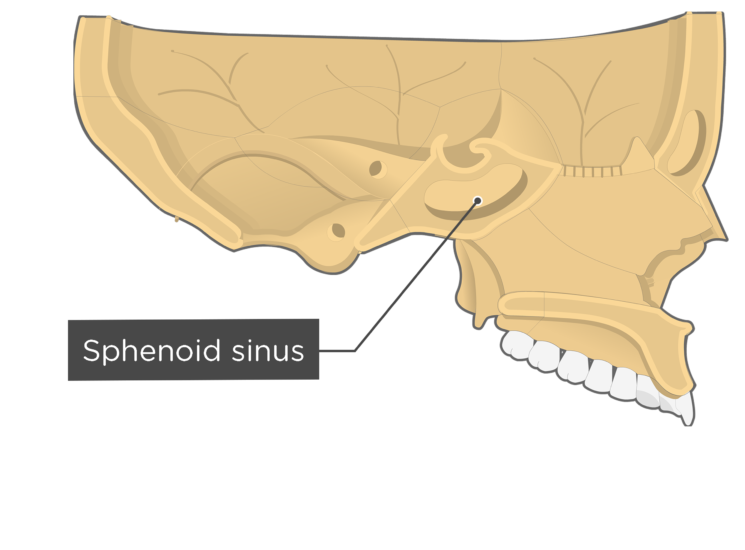
Sphenoid sinus anatomy. The sphenoid sinus is the most posterior paranasal sinus. The sphenoid sinuses are paired spaces formed within the body of the sphenoid bone. Paranasal air sinuses the sphenoidal sinuses are situated back of the nose in the sphenoidal bone which forms a forward part of the base of the skull and contains the depression or fossa for the pituitary gland.
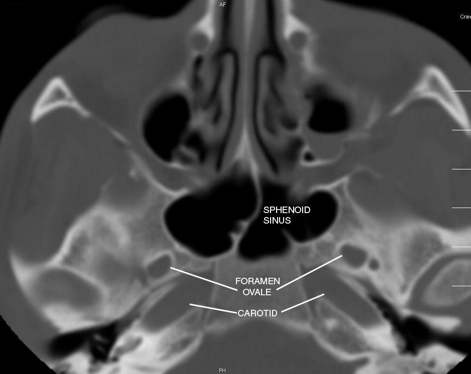
The sphenoid sinuses vary in size and shape and owing to the lateral displacement of the intervening septum which may insert on the carotid canal they are rarely symmetrical. Pneumatization starts at around 2 years of age and it develops more. The sphenoid sinuses are located in the sphenoid bone near the optic nerve and the pituitary gland on the side of the skull.
Significant variability exists regarding the dimensions of the sphenoid sinus attachment of the intersinus septum number of septa and pneumatization of the sphenoid bone. The apertures are high on the anterior walls of the sphenoid sinuses. They cannot be palpated during an extraoral examination.
There are seven bones that form the orbit eye socket and the sphenoid is one of these bones. This variability has a direct impact on surgical planning for endoscopic skull base cases. The sphenoid sinuses are highly variable in their configuration.
Normal anatomy variants. The sinuses are separated from each other by a bony wall or. Additionally pneumatization can also involve the posterior orbital wall pterygoid processes and lesser sphenoid wing.
The anatomy of the sphenoid sinus is highly variable. The sphenoid sinus is one of the four paired paranasal sinuses that is contained within the body of the sphenoid bone.
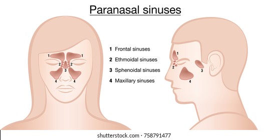
 Paranasal Sinuses
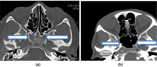 Incidental Discovery Of Sphenoid Sinuses Agenesis A Report
 Startradiology
 Figure 2 From The Maxillary Recess Of The Sphenoid Sinus
 Sphenoid Sinus Stock Photos Sphenoid Sinus Stock Images
 Ethmoid Sinus An Overview Sciencedirect Topics
Olfactory System Parts Function Organs Britannica
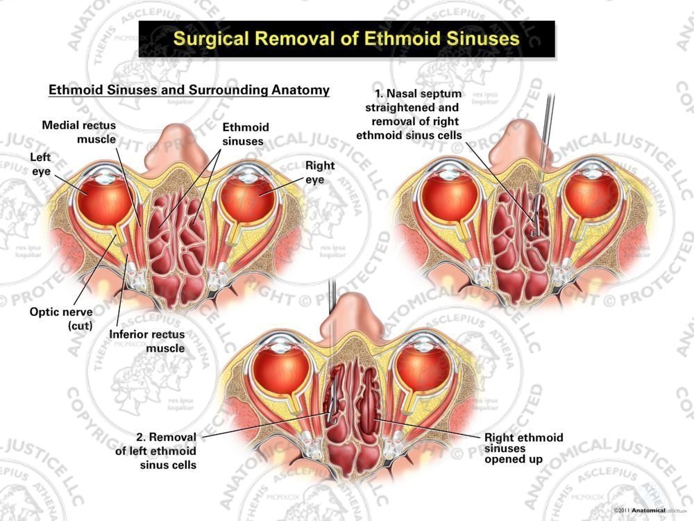 Surgical Removal Of Ethmoid Sinuses
 Anatomy Of Pns By Roohia


 Sphenoid Sinus With Nerves And Vessels Arteries Anatomy
 Nose And Sinus Anatomy Thomas S Higgins Md Msph
 Neurovascular Relationships Of The Sphenoid Sinus In
![]() Sphenoid Bone Anatomy Function And Development Kenhub
Sphenoid Bone Anatomy Function And Development Kenhub
 Paranasal Sinuses Wikipedia
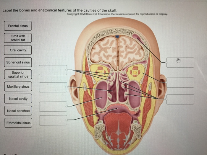 Solved Label The Bones And Anatomical Features Of The Cav
 Figure Anatomy Of The Cavernous Sinus Contributed By Okkes
 An Endoscopic View Showing Important Sphenoid Sinus Anatomy
 Ossicle Nose And Paranasal Sinuses Wikibooks Open Books
 Nose And Paranasal Sinus
 Definition Of Sphenoid Sinus Nci Dictionary Of Cancer
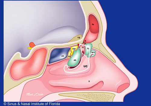 Sinus Nasal Institute Of Florida
 Sphenoidal Sinus An Overview Sciencedirect Topics
 Sphenoid Sinus Balloon Sinuplasty Medical Animation By Watermark
 Ethmoid Sinus
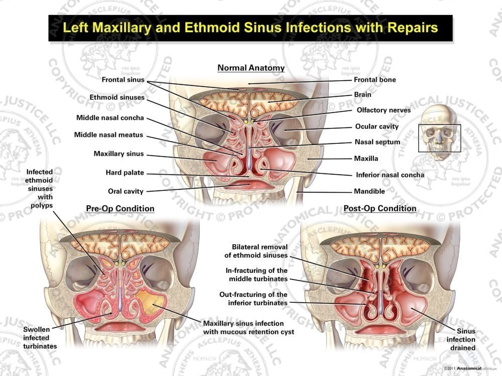 Left Maxillary And Ethmoid Sinus Infections With Repairs
![]() Paranasal Sinuses Anatomy And Clinical Aspects Kenhub
Paranasal Sinuses Anatomy And Clinical Aspects Kenhub
 Relationship Of Sphenoid Sinus With Hypophysis Cerebri 17
Belum ada Komentar untuk "Sphenoid Sinus Anatomy"
Posting Komentar