Anatomy Of A Model Cell
Distributes materials by diffusion. Cell theory first developed in 1839 by matthias jakob schleiden and theodor schwann states that all organisms are composed of one or more cells that cells are the fundamental unit of structure and function in all living organisms and that all cells come from pre existing cells.
 Anatomy And Physiology Parts Of A Human Cell
Learn vocabulary terms and more with flashcards games and other study tools.

Anatomy of a model cell. Anatomy of a model cell art labeling activity. Cell membrane barrier animation. The individual cell is the unit of structure of all living things.
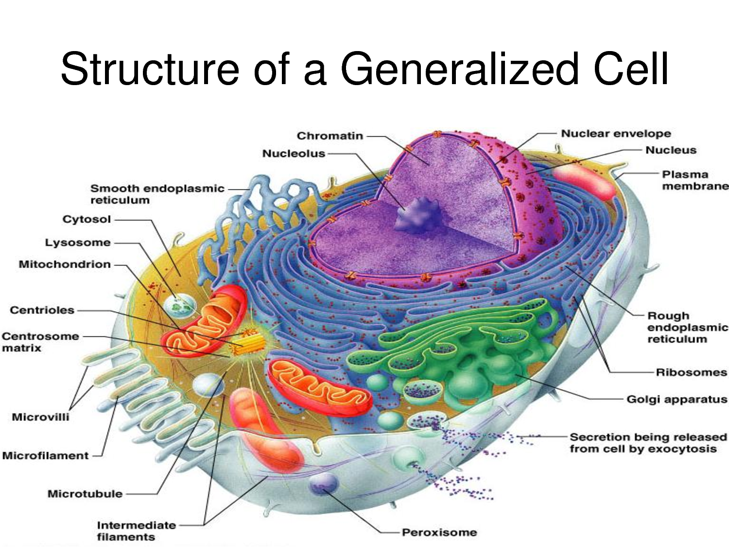
Some of the main organelles within the cell are the vacuoles mitochondria lysosomes ribosomes endoplasmic reticulum golgi apparatus and cell nucleus. Inside the cell is a jelly like fluid called cytoplasm that holds a cells organelles special structures that perform specific cell functions. The cytoplasm contains specialized organelles each of which is surrounded by a membrane.
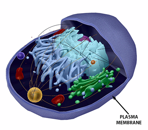
An entire organism may consist of a single cell which is called unicellular or many cells which is called multicellular. Composition of the plasma membrane lipid bilayer containing phospholipids steroids proteins and functions of the plasma membrane protection and support of the cell cells outer boundary. Chloroplasts convert light to chemical energy.
Start studying anatomy of a model cell. Plant cells differ from animal cells in that they lack centrioles and organelles for locomotion cilia and flagella but they do have additional specialized organelles. Within the cytoplasm of the cell are the organelles the cell requires to reproduce energy production and removal of waste.
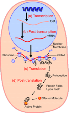
Centrioles the cytoplasm the rough and smooth endoplasmic reticulums the golgi complex lysosomes microfilaments mitochondria the nucleolus the nucleus the nuclear membrane pinocytotic vesicles the plasma membrane ribosomes and vacuoles. The nucleus art labeling activity. The cell contains a nucleus which contains the genetic material necessary for reproduction.
The anatomy of a model cell. The process of translation art labeling activity. Stages of a cells life cycle.
Anatomy of the plant cell. The quiz above includes the following features of a typical eukaryotic cell. Although animal cells dont have a cell wall they are protected by other cells such as white blood cells that fight disease.
 Trying To Study A Model Of The Cell Very Difficult Youtube
 Animal Cell Anatomy Enchantedlearning Com
 Fluid Mosaic Model Cell Membranes Article Article Khan
 Anatomy Wikipedia
 Plasma Membrane Of A Cell Definition Function Structure
 Cell Physiology Pathophysiology And The Relationship To All
 Novel Multiscale Insights Into The Composite Nature Of Water
The Cellular And Molecular Basis For Planarian Regeneration
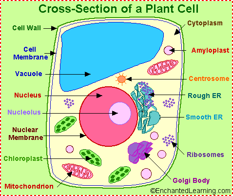 Plant Cell Anatomy Enchanted Learning
 Plant Cell Model Plant Cell Teaching Model Plastic Plant

 Smooth Er Definition Functions Structure Video

 Figure 4 From Anatomy Of A Blastocyst Cell Behaviors
 Chapter 3 The Cellular Level Of Organization Ppt Download
 2015 Pearson Education Inc Ppt Download
 3d Airway Cell Model Human Bronchial Epithelial Cells
 Animal Cell Model Diagram Project Parts Structure Labeled
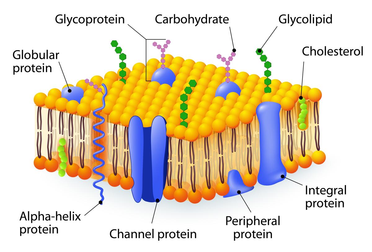 Fluid Mosaic Model
 Get The Cell S Anatomy Into A Cellbuilder
 Somso Model Of A Liver Cell
 Anatomy Of A Model Cell Diagram Quizlet
 Cell Biology Wikipedia
 Make A 3d Model Cell Cell Model Project Animal Cell
 Figure 3 From Anatomy Of A Blastocyst Cell Behaviors
Belum ada Komentar untuk "Anatomy Of A Model Cell"
Posting Komentar