Ankle Syndesmosis Anatomy
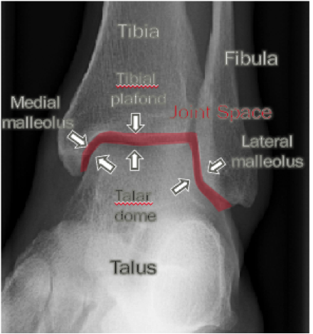
Anatomy the ankle is a synovial joint composed of the distal tibia and fibula as they articulate with the talus. Gross pt phd2 paul weinhold phd3 syndesmosis injuries are rare but very debilitating and frequently misdiagnosed.
 High Ankle Sprain Different Than Your Average Ankle Sprain
Only a few joints in the body are syndesmosis joints.
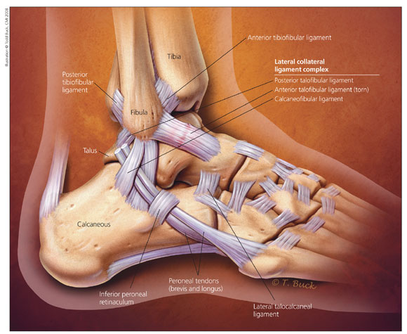
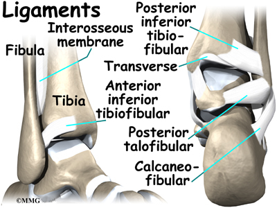
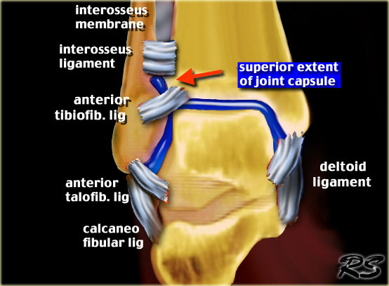
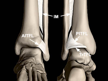
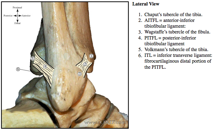
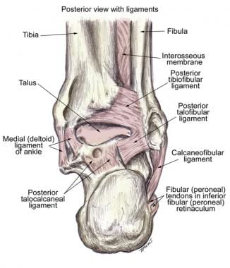
Ankle syndesmosis anatomy. The distal tibiofibular syndesmosis must be stable and congruent for normal ankle motion under physiological load. Ligaments distal tibiofibular syndesmosis includes anterior inferior tibiofibular ligaments aitfl originates from anterolateral tubercle of tibia chaputs inserts on anterior tubercle of fibula wagstaffes posterior inferior tibiofibular ligament pitfl originates from posterior tubercle of tibia volkmanns inserts on posterior part of. In addition to the ankle syndesmosis the connection of the tibia and fibula syndesmosis joints are also located in the lower spine where the top of the triangular shaped sacrum bone fits between the pelvis bones.
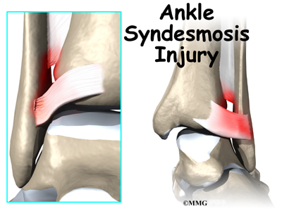
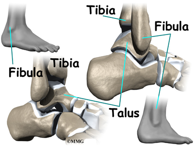
Syndesmotic injury usually occurred in ankle sprain with external rotation of the talus that resulted at either ligamentous rupture or bony avulsion of the syndesmotic ligament complex. Distal tibiofibular syndesmosis injuries are a relatively frequent ankle injury although less common than a fracture or lateral collateral ligament injuries. The distal tibia and fibula articulate with each other at the distal tibiofibular joint which is more commonly referred to as the tibiofibular syndesmosis or simply the syndesmosis.
The superior or proximal tibiofibular joint the interosseous membrane and the inferior or distal tibiofibular joint. However there are three definable ligaments at the ankle as follows. This articulation of the fibula with the tibia can be subdivided further into 3 regions.
It is reported ankle syndesmosis injury occurs in 1 11 of all ankle sprains without fractures however 40 of patients report ankle instability six months post ankle sprain. A third articulation in the region of the ankle and lower leg is between the tibia and fibula. Injuries can occur with any ankle motion but the most common motions are extreme external rotation or dorsiflexion of the talus.
A syndesmotic or high ankle sprain is one that involves the ligaments binding the distal tibia and fibula at the distal tibiofibular syndesmosis. Anatomy biomechanics mechanism of injury and clinical guidelines for diagnosis and intervention cheng feng lin ms1 michael t. The syndesmosis of the ankle refers to the membrane connecting the tibia to the fibula.
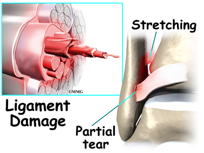
The tibia and fibula are connected throughout their length by an interosseous membrane. They are estimated to comprise 10 range 1 20 of ankle injuries. The instability could result from widening of the ankle mortice following stretching of the ligaments.
 High Ankle Sprain Ankle Syndesmosis Eorthopod Com
 The Radiology Assistant Ankle Mri Examination
 High Ankle Sprain Ankle Syndesmosis Eorthopod Com
 Anatomy Of The Ankle Maxeffortmuscle Com
High Ankle Sprain Vs Ankle Sprain What S The Difference
 Not All Ankle Sprains Are Created Equal Nuem Blog
 Anatomy 101 Ankle Syndesmosis Distal Tibiofibular Joint
 Not All Ankle Sprains Are Created Equal Nuem Blog
 X Enkel Startradiology
 Not All Ankle Sprains Are Created Equal Nuem Blog
![]() High Ankle Sprain Syndesmosis Injury Foot Ankle
High Ankle Sprain Syndesmosis Injury Foot Ankle
 The Radiology Assistant Ankle Fracture Mechanism And
 Syndesmosis An Overview Sciencedirect Topics
 Ankle Anatomy Eorif
 Anatomical Illustration Of The Tibiofibular Syndesmosis
 Ankle Joint Anatomy Overview Lateral Ligament Anatomy And

![]() Ankle Joint Anatomy Bones Ligaments And Movements Kenhub
Ankle Joint Anatomy Bones Ligaments And Movements Kenhub
 Physical Therapy In Plymouth For Ankle Pain Anatomy
 High Ankle Sprains Syndesmosis Injury Footnotes Publishing
Belum ada Komentar untuk "Ankle Syndesmosis Anatomy"
Posting Komentar