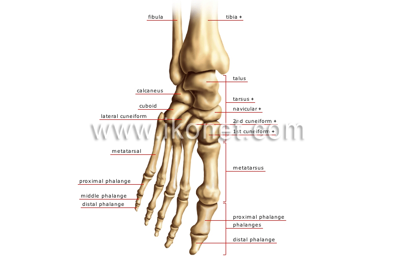
Cuneiform Anatomy
The intermediate cuneiform second cuneiform or middle cuneiform is shaped like a wedge. The medial cuneiform is one of the cuneiforms it is the most medial in the distal row of tarsal bones.
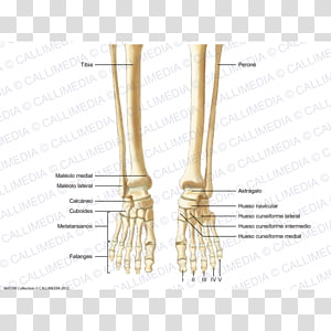
 Foot Bones Diagram Quizlet
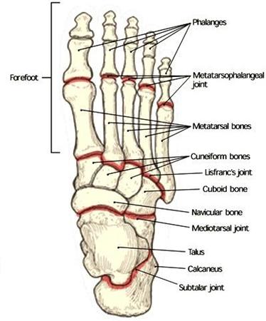
There are three cuneiform bones medial intermediate and lateral.

Cuneiform anatomy. The medial cuneiform is the largest and the intermediate cuneiform the smallest. There are three cuneiform bones. In the medial cuneiform the edge of the wedge forms the dorsal surface.
We think this is the most useful anatomy picture that you need. It has a narrow dorsal surface and a flat plantar surface which receives a slip from the tibialis posterior tendon. Medial first cuneiform bone intermediate middle second cuneiform bone lateral third external cuneiform bone.
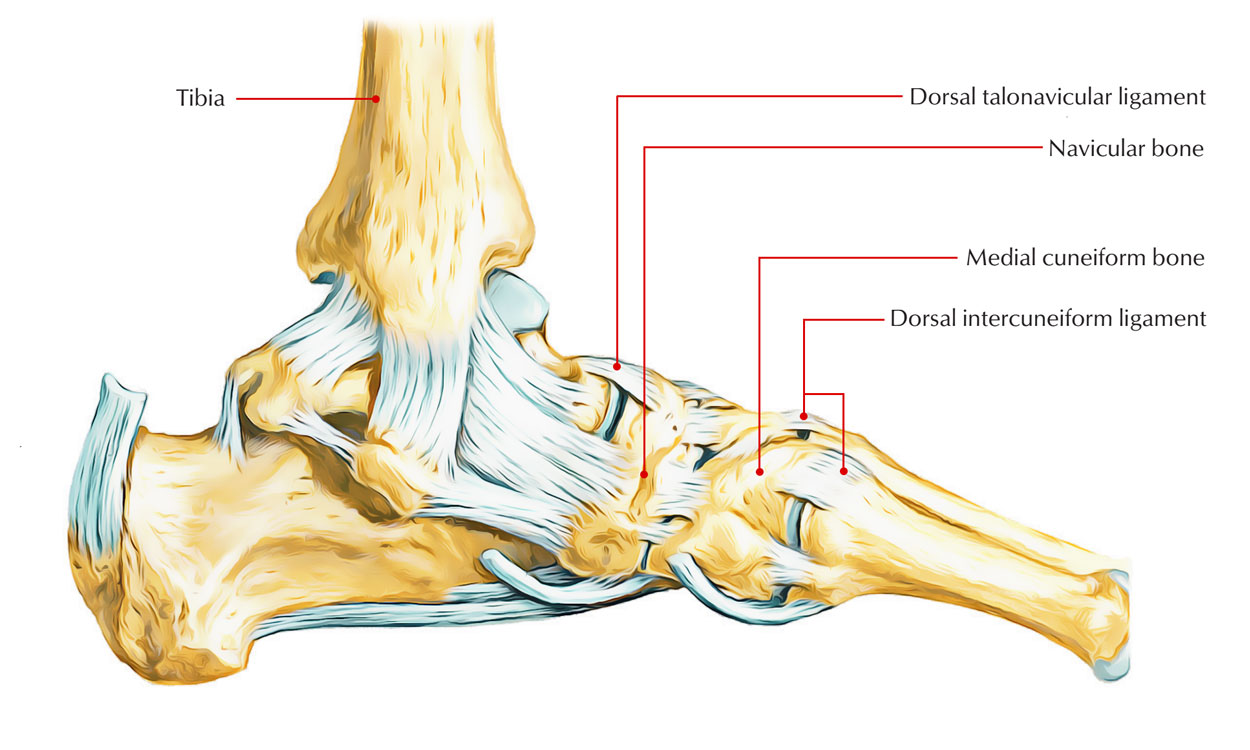
The distal surface is reniform congruent with the articulating base of first metatarsal and proximal surface has a pyriform facet for the navicular. The medial cuneiform is the largest of the three cuneiform bones. We think this is the most useful anatomy picture that you need.
The medial cuneiform also known as first cuneiform is the largest of the cuneiforms. The three cuneiform bones latin. As their name suggests these are wedge shaped bones.
The lateral cuneiform occupies the center of the front row of the tarsal. The lateral cuneiform also known as third cuneiform or external cuneiform. The intermediate cuneiform bone is the smallest of the three cuneiform bones.
A wedge shaped bone especially any of the three bones located in the tarsus of the foot. Cuneiform bones medial cuneiform. Ossa cuneiformia are located between the navicular bone and the first three metatarsal bones and medial to the cuboid bone.
The medial cuneiform participates in articulation joint movement along with the first and second metatarsals the intermediate cuneiform and the navicular. The bone functions as the attachment for numerous ligaments fibrous connective tissue such as those of peroneus longus and the tibialis anterior muscles. The three cuneiforms are named.
The Medial Cuneiform Bone Stock Illustration K28860039
![]() Cuneiform Bones Anatomy And Clinical Notes Kenhub
Cuneiform Bones Anatomy And Clinical Notes Kenhub
 Cuneiform Bone An Overview Sciencedirect Topics
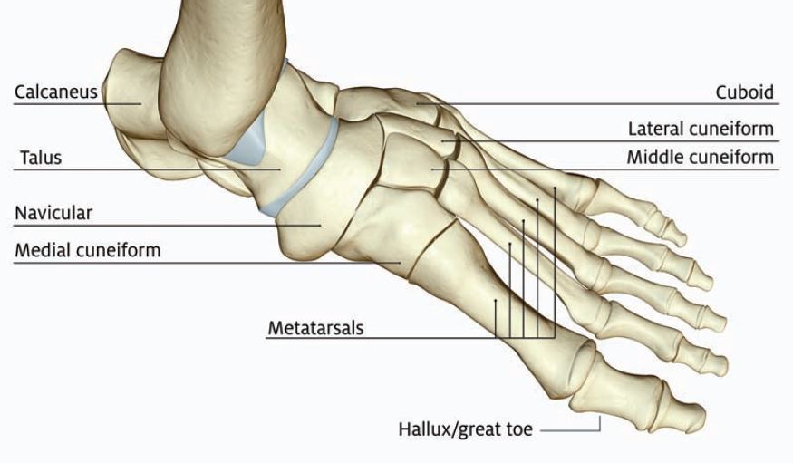
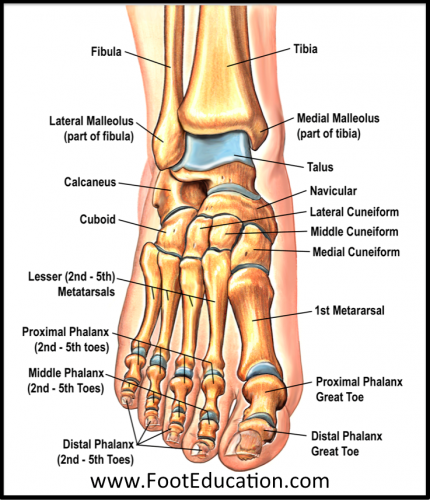 Bones And Joints Of The Foot And Ankle Overview Footeducation
 Cuneiform Anatomy Wikipedia The Free Encyclopedia Toe
 Cuneiform Bones Transparent Background Png Cliparts Free
 Cuneiform Bones Wikipedia
 Medial Cuneiform Radiology Reference Article Radiopaedia Org
 The Medial Cuneiform Bone
 Cuneiform Orthopaedicsone Review Orthopaedicsone
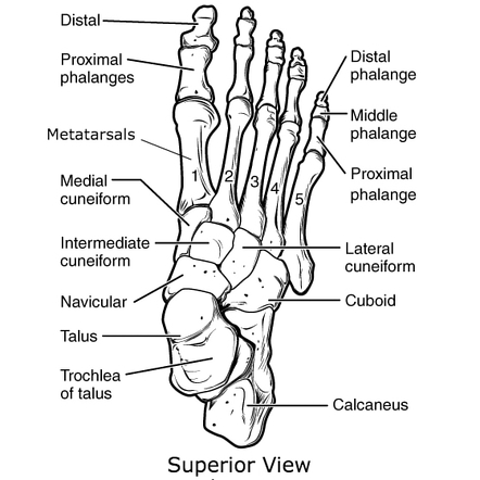
Intermediate Cuneiform Middle Cuneiform
 Anatomical Location Of The Cuneiform Nucleus Download
 Tarsometatarsal Intermetatarsal Articulations Prohealthsys
 Lisfranc Injury Tarsometatarsal Fracture Dislocation
 Bones Of The Foot Cuneiform Bones Human Anatomy Kenhub
 Dorsal View Of The Skeletal Anatomy Of The Right Foot
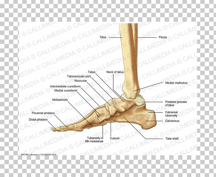
 Duke Anatomy Lab 2 Pre Lab Exercise
 Easy Notes On Navicular Bone Learn In Just 4 Minutes
 Foot Anatomy And Biomechanics Foot Ankle Orthobullets
 Managing Foot Fractures In Urgent Care Journal Of Urgent
 Tarsal Bones Anatomy Skeletal System Review
![]() Cuneiform Bones Anatomy And Clinical Notes Kenhub
Cuneiform Bones Anatomy And Clinical Notes Kenhub
 Medial Cuneiform Bone Stock Photos Medial Cuneiform Bone
 Finger Foot Cuneiform Bones Medial Cuneiform Bone Anatomy
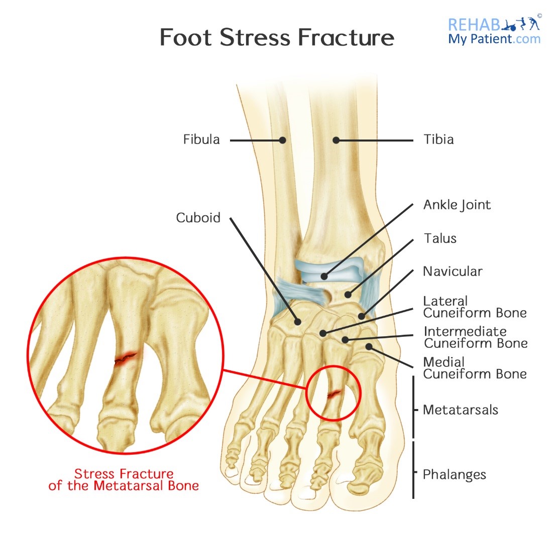 Foot Stress Fracture Rehab My Patient
![]() Cuneiform Bones Anatomy And Clinical Notes Kenhub
Cuneiform Bones Anatomy And Clinical Notes Kenhub


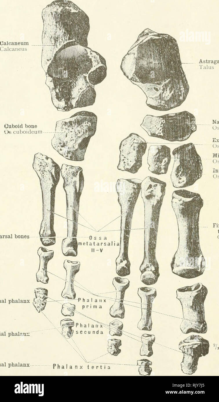
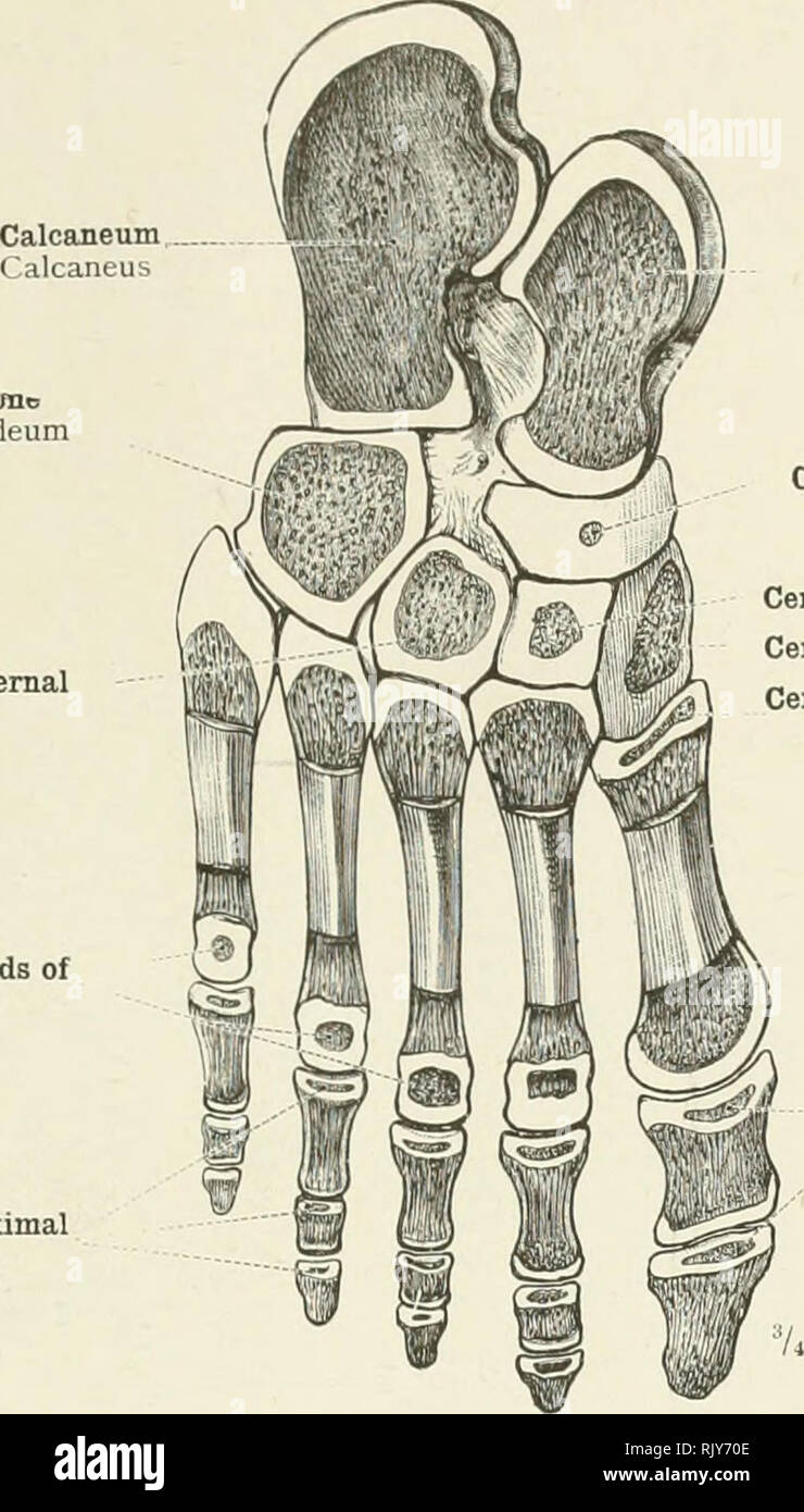 An Atlas Of Human Anatomy For Students And Physicians
Belum ada Komentar untuk "Cuneiform Anatomy"
Posting Komentar