Pelvis Mri Anatomy
Mri provides superior soft tissue contrast resolution for imaging the anatomy best seen in t1 weighted and pathology best seen on t2 weighted of the pelvis 3. The bony pelvic girdle consists of the innominate bones bilaterally and the sacrum and coccyx posteriorly.
 Normal Female Pelvis Axial A And Sagittal B Mri T2
Anatomy of the female pelvis mri atlas of the human body using cross sectional imaging.

Pelvis mri anatomy. This muscle originates from the inferior ramus of the pubic bone and attaches to the gluteal tuberosity and adductor tubercle of the femur. The multiplanar capabilities and excellent soft tissue contrast on magnetic resonance imaging mri of the pelvis provide superb depiction of the female pelvic anatomy and often lead to specific diagnosis without ionizing radiation. The pelvic diaphragm is composed of the ischiococcygeus muscle and levator ani muscle the latter of which consists of the iliococcygeus puborectalis and pubococcygeus muscles.
Mri of the male pelvis. 7 gluteus maximus m. Figure 3b schematics show the anatomy of the female pelvic floor at the level of the pelvic diaphragm a and the urogenital diaphragm b.
Use the mouse scroll wheel to move the images up and down alternatively use the tiny arrows on both side of the image to move the images. Mri is often used as a problem solving tool in patients where ultrasound is inconclusive or suboptimal. Because it has two heads both an adductor and hamstring part.
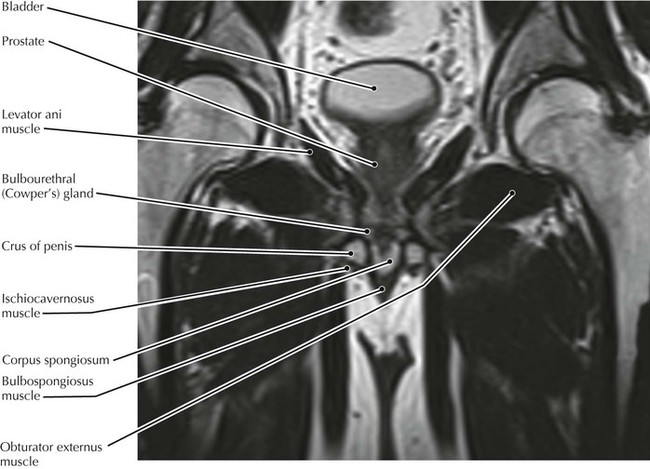
Knee shoulder shoulder arthrogram ankle elbow wrist hip contact. In an adult the innominate bones consist of the fused ilium ischium and pubis figure 1. 1 corpus cavernosum 2 corpus spongiosum bulb of the penis 3 ramus ischium 4 ischiocavernosus m.
5 anal canal 6 sphincter ani externus m. This mri male pelvis axial cross sectional anatomy tool is absolutely free to use. Anatomy of the pelvis bony anatomy.
Magnetic resonance mr imaging is a valuable technique for the non invasive evaluation of the female pelvic region for example diagnosing or staging developmental anomalies leiomyomas adenomyosis vaginal neoplasms endometrial or cervical carcinoma. Use the mouse scroll wheel to move the images up and down alternatively use the tiny arrows on both side of the image to move the images. This mri female pelvis sagittal cross sectional anatomy title tool is absolutely free to use.
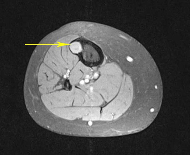
47 adductor magnus muscle this is the largest of the group of adductors of the thigh. Atlas of mri of the male pelvis. Mri of the male pelvis.
Angiography invasive angiography is the gold standard modality for assessing pelvic vasculature 3. Use the mouse to scroll or the arrows.
 X Rays Ct Scans And Mris Orthoinfo Aaos
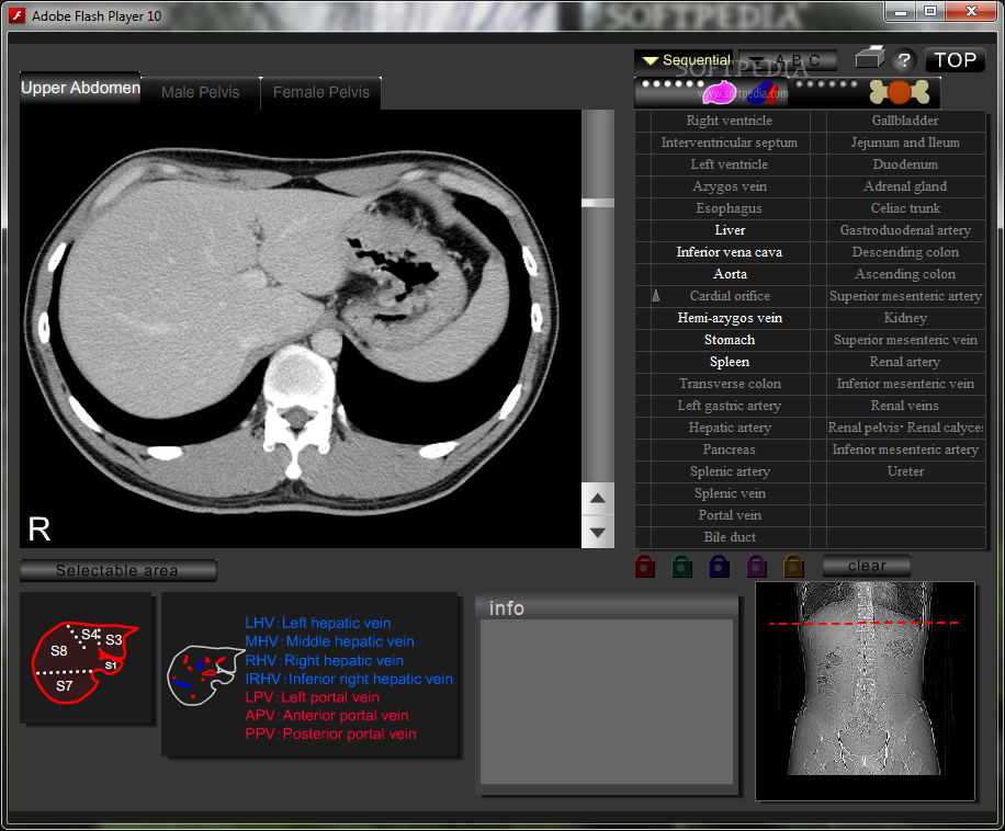 Download Ct And Mri Interactive Atlas Of Cross Sectional Anatomy 1 1e
 Ecr 2013 C 1484 Mri And Us Anatomy Of Female And Male
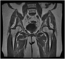
 Hip Mri Approach To Msk Mri Series
 Hip Mri
 Mri Anatomy Of The Hip Review Mri Anatomy Of The Hip
 Mri Pelvis Of A Woman Benigne Tumor Uterine Myoma Weight
 Mri Based Detailed Evaluation Of The Anatomy Of The Human
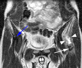 Apophyseal Avulsion Injury Of The Pelvis Radsource
 Atlas Of Mri Of The Female Pelvis
 Pelvic Mri For Fibroids Why You Need An Mri To Detect Fibroids
 07 Mri Femal Pelvis
 Mri Pelvis Anatomy Free Male Pelvis Axial Anatomy
 Anatomy Of The Female Pelvis The Bmj
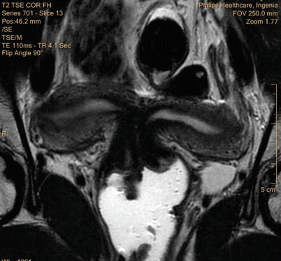
 Mri Pelvis
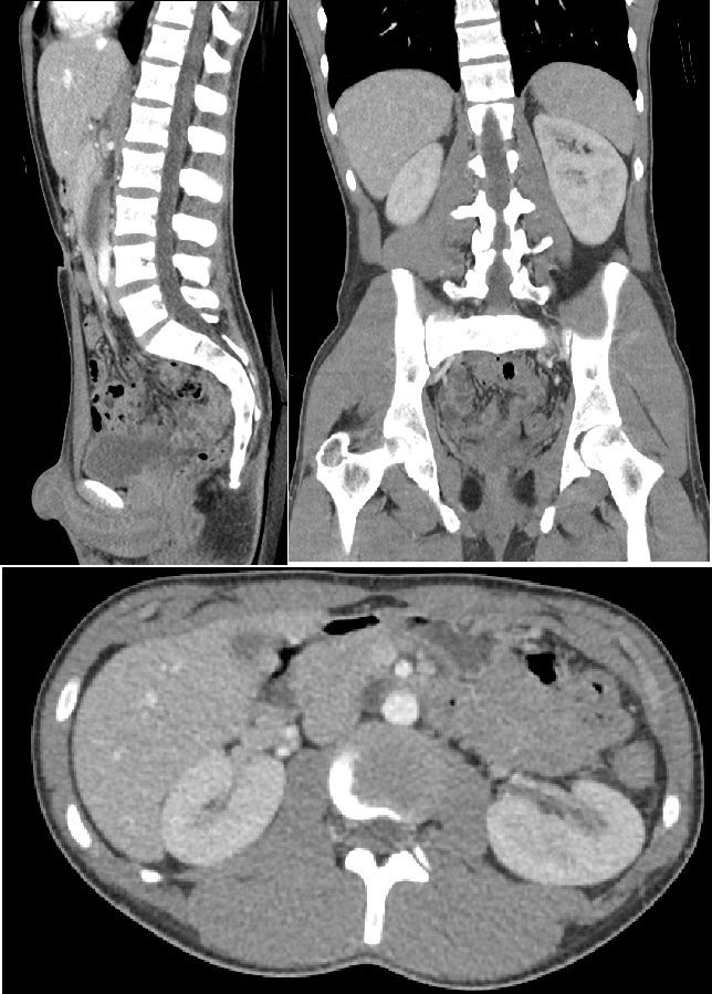 Computed Tomography Of The Abdomen And Pelvis Wikipedia
Mri Pelvis Anatomy Free Male Pelvis Axial Anatomy
 Atlas Of Mri Of The Male Pelvis
 Mri Pelvis Anatomy Free Male Pelvis Axial Anatomy
 Getting Ready For An Mri Of Your Pelvis Sansum Clinic
 Benign Disease Of The Uterus Springerlink
 Atlas Of Mri Of The Female Pelvis
 Radiology Images
 Sectional Anatomy Quiz 7 Pelvis Flashcards Quizlet
 Figure 1 From Does Bilateral Sacrospinous Fixation With
 Presentation1 Pptx Radiological Anatomy Of The Abdomen And
 The Hip Anatomy On 3t Mr And 3d Pictures
 Normal Gynecologic Anatomy
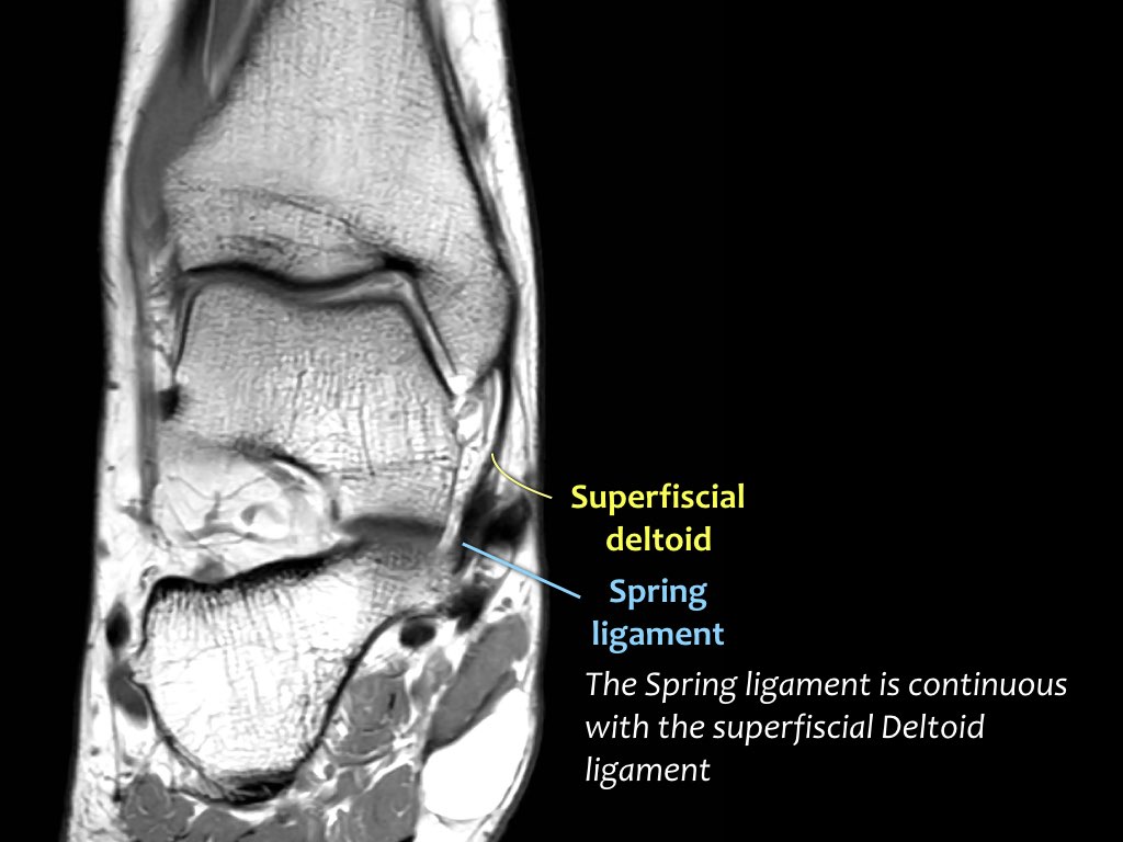 The Radiology Assistant Ankle Mri Examination
 Pelvis And Perineum Radiology Key
 The Male Pelvis Mr Anatomy Atlas Of The Prostate Bladder
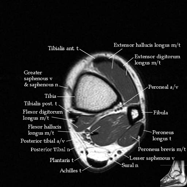 Mri Ankle Anatomy
 Module 5 Pelvis Imaging
 Pelvic Mri Showing A Large Uterus With Disappearance Of Its

Belum ada Komentar untuk "Pelvis Mri Anatomy"
Posting Komentar