Deltoid Anatomy
Use the mouse to scroll or the arrows. Knee shoulder shoulder arthrogram ankle elbow wrist hip contact.
 Deltoid Muscle Wikipedia
The shoulder joint is formed where the humerus upper arm bone fits into the scapula shoulder blade like a ball and socket.

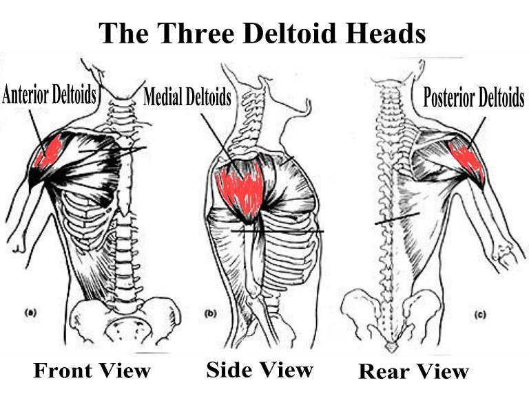
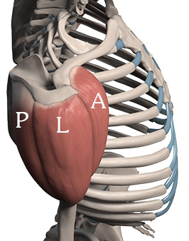
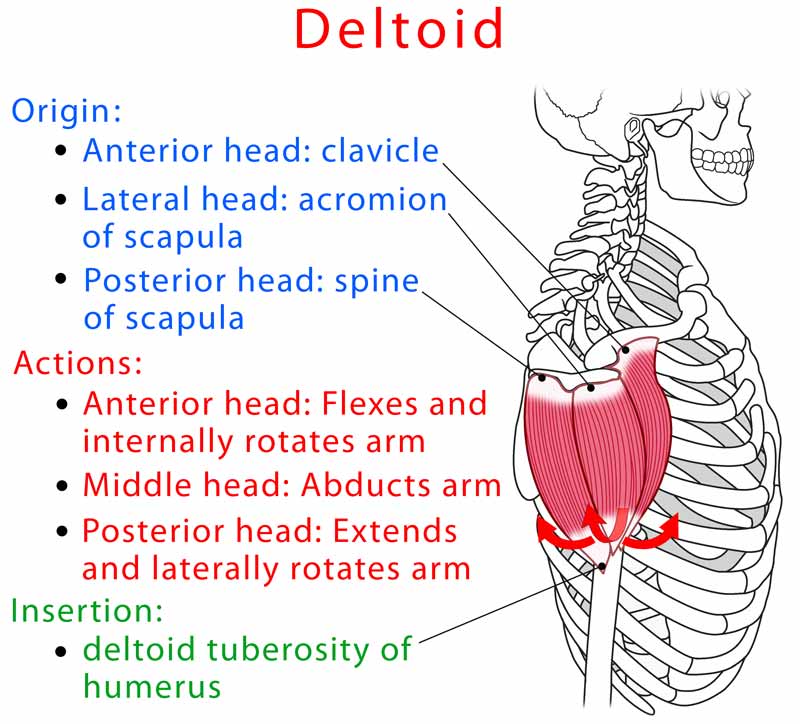
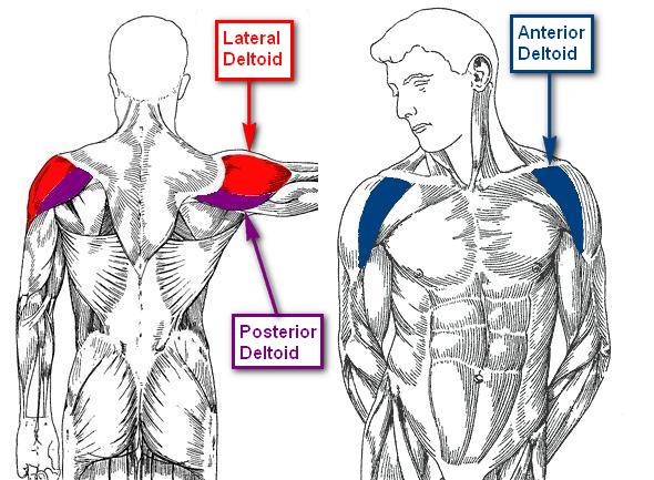
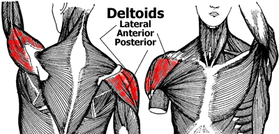
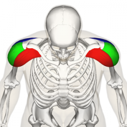
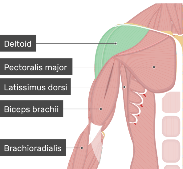
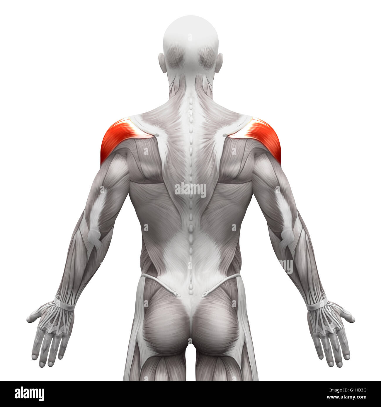
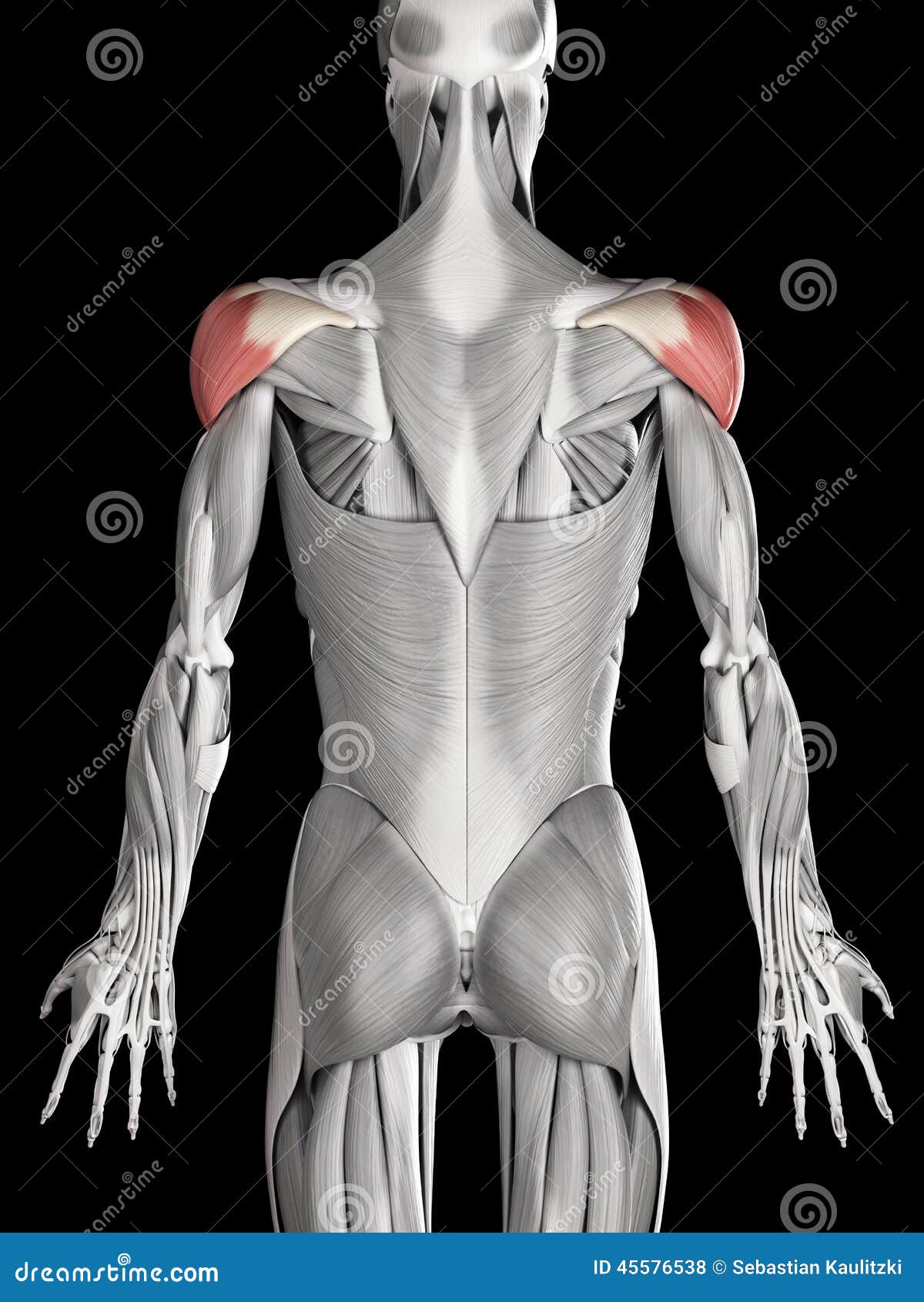
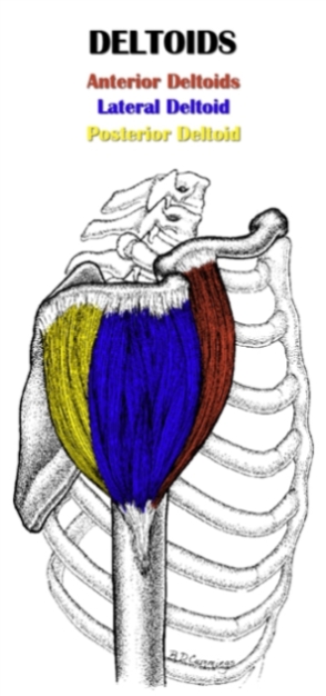
Deltoid anatomy. The shoulder is a complex combination of bones and joints where many muscles act to provide the widest range of motion of any part of the body. Previous studies showed that the insertion of the intramuscular tendons of the deltoid muscle formed three discrete sets of muscle fibers often referred to as heads. The deltoid is attached by tendons to the skeleton at the clavicle collarbone scapula shoulder blade and humerus upper arm bone.
Cause learning muscle anatomy can be fun. Anatomy of the deltoid muscle. In this video the followi.
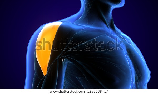
The glenohumeral joint is where the ball humeral head and the socket the glenoid meet. Numerous muscles help stabilize the three joints of. The deltoid muscle is a large and powerful muscle of the shoulder joint.
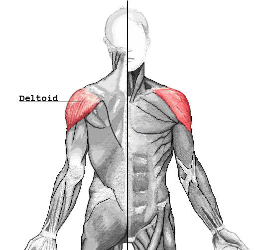
It distinctively shapes the surface anatomy of the shoulder and this muscle is supplied by the axillary nerve a branch of the brachial plexus. It is named after the greek letter delta which is shaped like an equilateral triangle. The shoulder is made up of two joints the acromioclavicular joint and the glenohumeral joint.
The shoulder is one of the largest and most complex joints in the body. Learn about the deltoid its origin insertion and action and all the fun facts in between. The deltoid muscle is a rounded triangular muscle located on the uppermost part of the arm and the top of the shoulder.
Lateral or acromial fibers arise. The acromioclavicular joint is where the acromion part of the shoulder blade scapula and the collar bone clavicle meet. The anterior or clavicular fibers arise from most of the anterior border and upper surface.
The anatomy of the shoulder. This may cause medical professionals to mistake the separate muscle fibers of the posterior deltoid for an adjoining muscle teres minor. A common anatomical variation of the deltoid includes the presence of separate fascial sheaths and muscle fibers on the posterior deltoid.
And with new paints this time.
![]() Deltoid Muscle Anatomy Function And Clinical Aspects Kenhub
Deltoid Muscle Anatomy Function And Clinical Aspects Kenhub
 Bodybuilding Anatomy Shoulders Bodybuilding Wizard
 How To Draw Deltoids Anatomy For Artists Proko
 Deltoid Front Lateral Rear Anatomy Location Function
 Exercises To Develop Monstrous Delts
 The Deltoid Muscle Get The Basic Facts About It
How To Stretch The Anterior Deltoid Anatomy
 Deltoid Muscle Origin Insertion Action Human Anatomy Kenhub
 Deltoid Wiktionary
 Deltoid Muscle Anatomy For Positioning Of Ultrasound Probe
 Paint Draw Paint Learn To Draw Anatomy Basics The Deltoid
 What Does The Deltoid Muscle Do Definition Function
 Deltoid Muscles Anatomy Map
 Drawing Deltoid Muscles Anatomy Map Clipart Drawing
 Image Result For Shoulder And Deltoid Anatomy Bones And
 Lateral Deltoid Functional Anatomy Guide Bodybuilding Wizard
 Shoulder Training The Best Exercises For The Deltoid
 Anterior Deltoid Functional Anatomy Guide Bodybuilding Wizard
 Deltoid Trigger Points And Referred Pain Patterns
 Deltoid Physiopedia
 3d Illustration Shoulders Deltoid Anatomy Muscles Stock
 Deltoid Muscle Anterior And Middle Heads
 Anatomy Deltoid Shoulder Muscle Anatomy Shoulder Anatomy
 Scapula Approach Deltopectoral Approach Ao Surgery


 Deltoid Muscle Anatomy Muscles Isolated On White 3d
 Hsf1 Exam 2 Anatomy Deltoid 2 Diagram Quizlet
 Pin On Writing
 The Deltoid Stock Illustration Illustration Of Anatomy
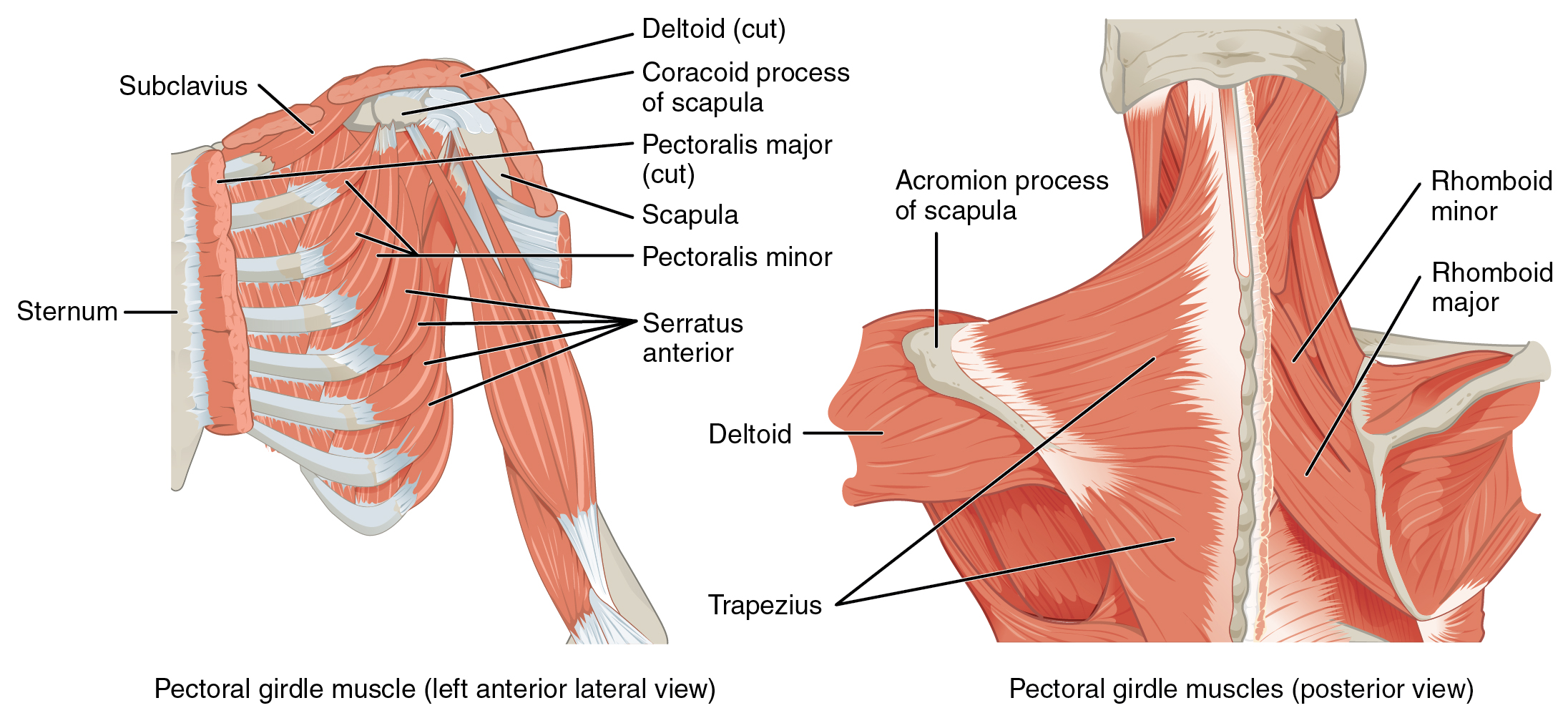 11 5 Muscles Of The Pectoral Girdle And Upper Limbs
 The Ultimate Anterior Deltoid Anatomy Exercise Training Guide
Belum ada Komentar untuk "Deltoid Anatomy"
Posting Komentar