Horse Foot Anatomy
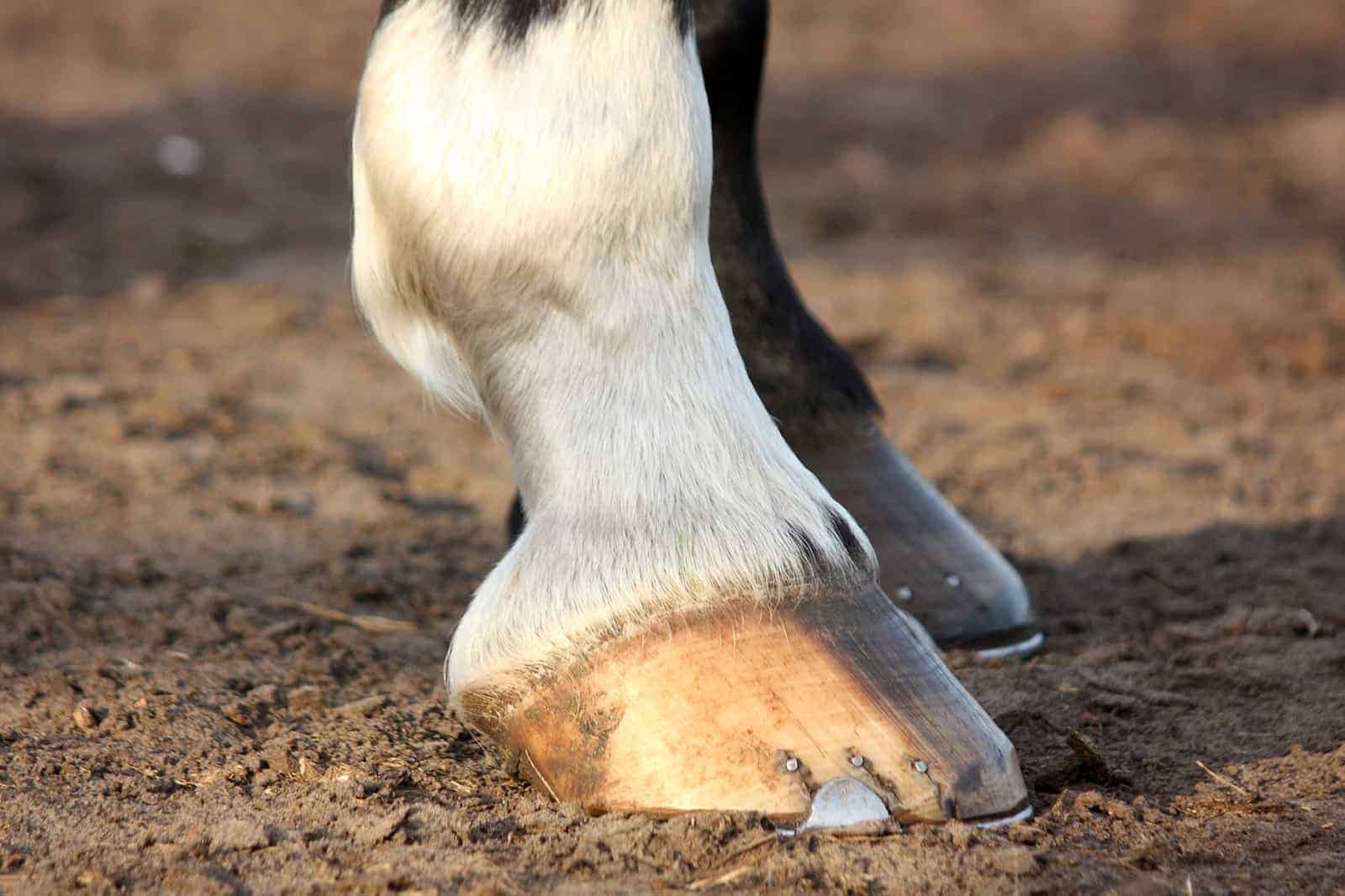
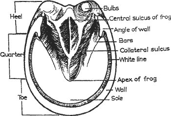
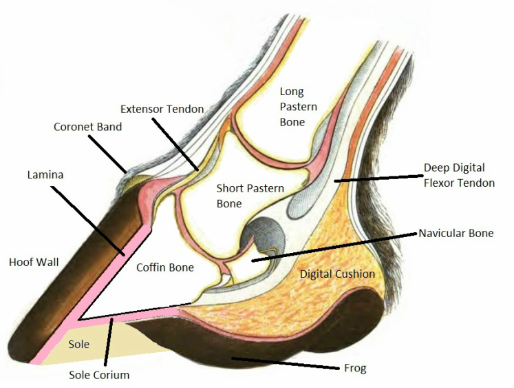
From equine skeletal anatomy to body parts and teeth. When the foot is lifted off the ground the sole and frog are visible as well as the bars of the wall and the collateral grooves figure 1.
 Hoof Anatomy A Beginner S Guide The Equine Podiatry
Outside underside and inside.
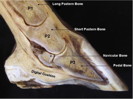
Horse foot anatomy. This is why keeping a horse in a stall with little activity is so unhealthy. By just observing the feet you can learn a lot about the horse. It covers the front and sides of the third phalanx or coffin bone.
Cartilage extends backwards and upwards from this bone. In part this is a result of solar concavity which has a variable depth in the region of 115 cm. Diagrams illustrations and charts will help you understand how your horse is put together.
Get the basics on horse anatomy that every horse owner needs. Just squeezing the heels by hand will demonstrate that. New hoof layers grow just beneath the coronet.
The horses hoofs are an amazing structure. Also called third phalanx the coffin bone is the lowest in the horses foot connecting to leg muscles via tendons. Its made up of several different parts all serving a different purpose yet working in symmetry to keep the horse sound and healthy.
Functional anatomy of the horse foot. The wall is made up of the toe front quarters sides and heel. It is elastic and flexible.
The hoof can be broken into three different areas for better understanding of its structures and purposes. Just pick an audience or yourself and itll end up in their incoming play queue. The horse hoof anatomy allows the hoof to act as a second heart.
Home about equine podiatry articles hoof anatomy a beginners guide the horses hoof is a miracle of engineering. 0 0000 a shoutout is a way of letting people know of a game you want them to play. What horse hooves are made of.
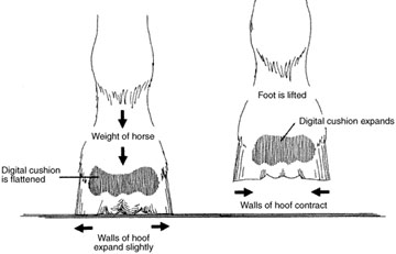
When loaded the hoof physiologically changes its shape. It contains a whole host of structures which when healthy operate in equilibrium with each other to form a hoof capsule which is able to withstand huge forces utilising energy to assist with forward movement while providing protection to the sensitive structures beneath. General health and the horses environment will leave tell tale signs on his hooves.
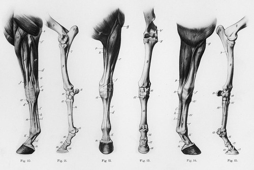
Develop a better understanding of where leg injuries occur and the inner workings of the horse hoof. You can see evidence of his conformation and how he moves by the way that his feet grow and wear. Coronary band the point where the skin and hair meets the hoof wall.
The horse hoof is not at all a rigid structure. With each step the horse takes the flexible hoof expands and contracts forcing blood back up the horses leg toward the heart.
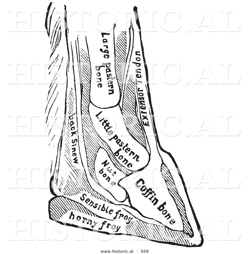
 Farriery Seventeen Figures Including The Anatomy Of Horses
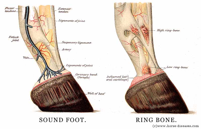
 An Overview Of The Inferior Check Ligament In Horses
 Hoof Care Tips And Anatomy
 Causes Of Equine Lameness Equimed Horse Health Matters
Horse Hoof Anatomy A Guided Tour The Horse
 Contraction Continued Modern Blacksmithing 1901
 Treating Soft Tissue Injuries American Farriers Journal
 No Hoof No Horse Uf Ifas Extension Marion County
 Farriery Anatomy Of The Horse S Hoof And Shoes Etc
 Horse Hoof Anatomy Teaching Chart
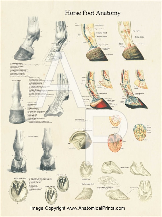 Horse Foot Hoof Veterinary Anatomy Poster 18 X 24
 A Working Knowledge Of Anatomy Is Important To Everyday
 Hoof Foot Anatomy Of The Horse Print Art Print By Aburnski
 Educational Horse Hoof Anatomy Information
 Horse Anatomy Mobility Health
 Historical Vector Illustration Of A Horse Diagram Featuring
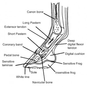 Anatomy Of The Leg And Hoof Vikki Fear Dep Mepa Uk
 Everything You Need To Know About Laminitis Irongate
 What Is Horse Laminitis And How To Deal With Easy And Severe
 Limbs Of The Horse Wikipedia
 Laminitis Not Just A Risk For Fat Ponies Vet In
 The Anatomy And Physiology Of The Hoof Australia Life
 Anatomy Horse Foot Hoof Picture
 Horse Hind Foot With Hoof Natural Specimen Anatomy Model Articulated


Belum ada Komentar untuk "Horse Foot Anatomy"
Posting Komentar