Lung Heart Anatomy
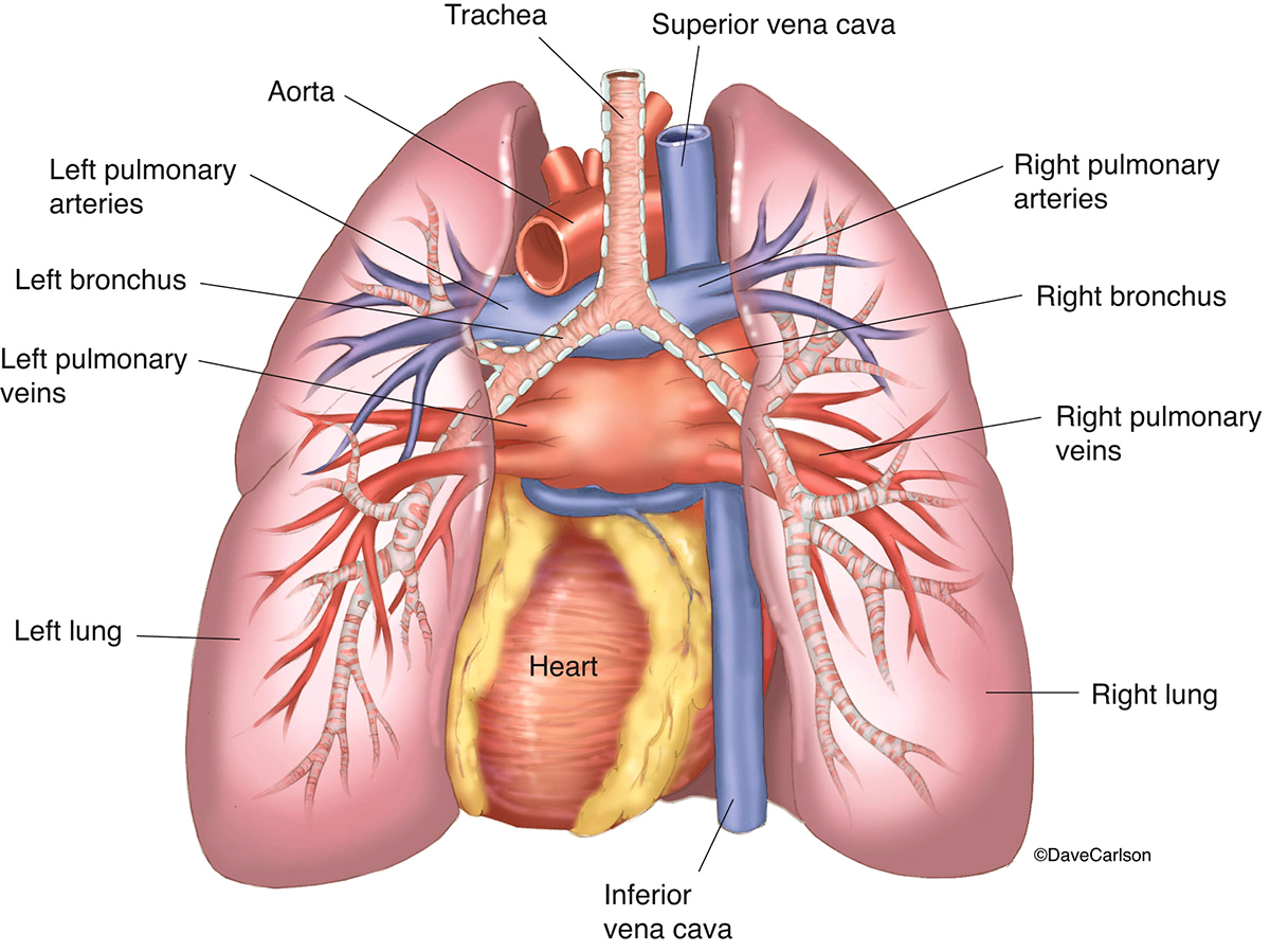
Left superior lobar bronchus 5. Lung anatomy this spongy pinkish organ looks like two upside down cones in your chest.
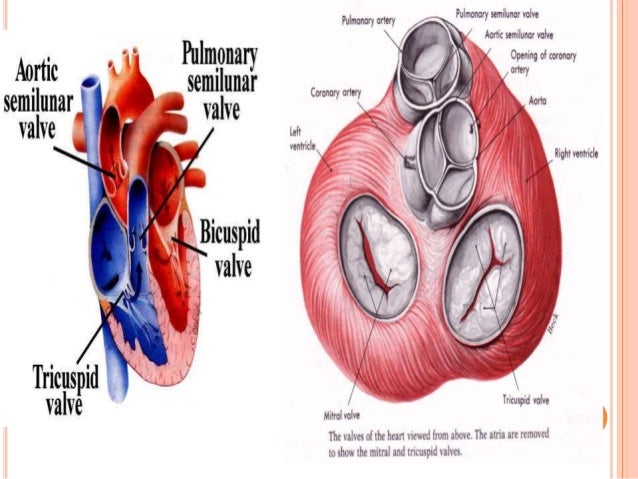 Anatomy And Physiology Of Heart Lung
For a random human anatomy.

Lung heart anatomy. The lungs are covered by a thin tissue layer called the pleura. Surfaces three these correspond to the area of the thorax that they face. Lobes fissures surfaces their shapes and stuff like that.
Anatomy and physiology of heart lung thoracic cavity bloodvessels. The same kind of thin tissue lines the inside of the chest cavity also called pleura. The right lung has three lobes.
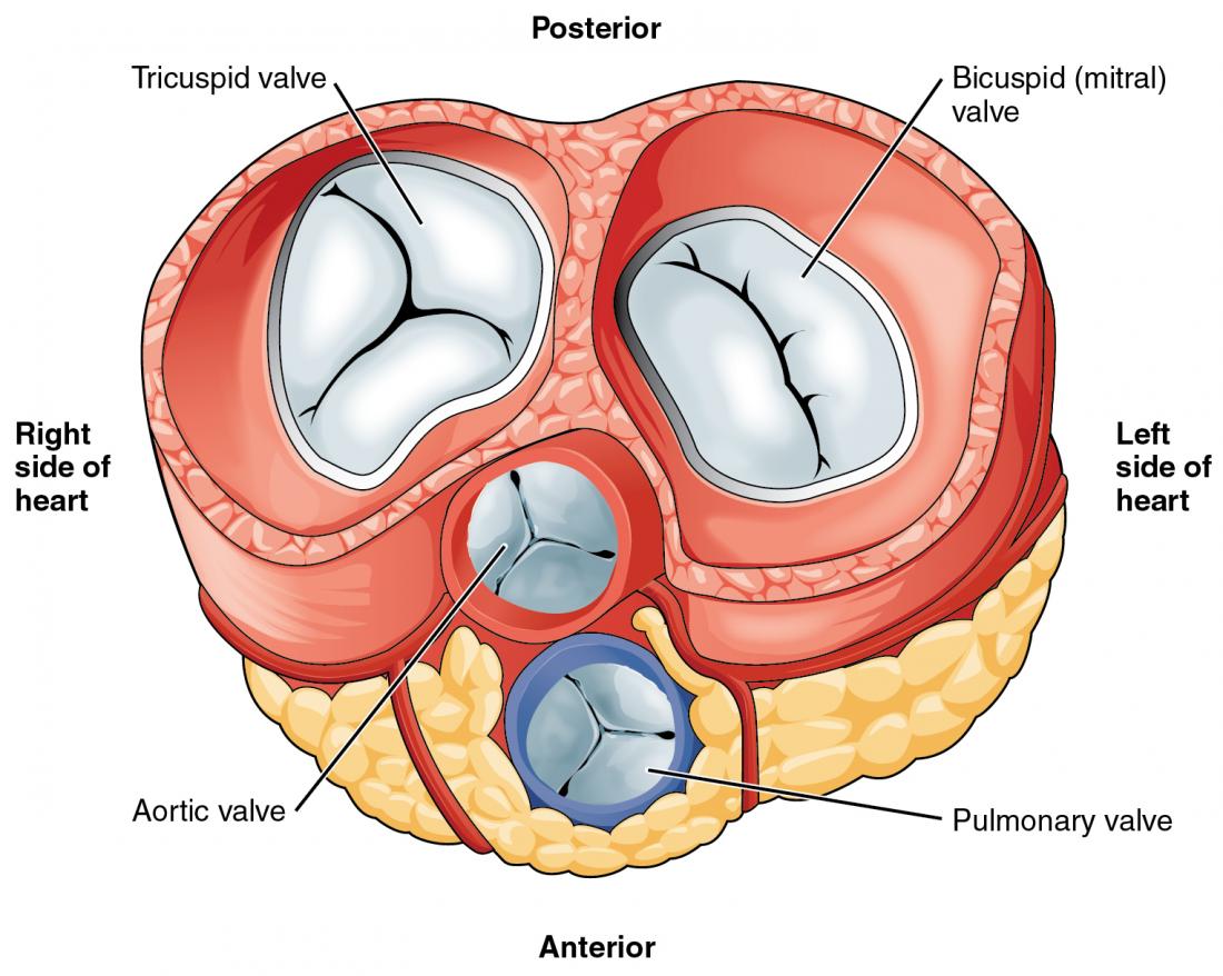
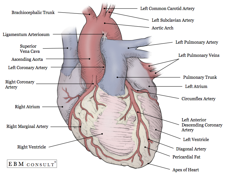
The left lung has only two lobes to make room for your heart. The human heart is located within the thoracic cavity medially between the lungs in the space known as the mediastinum. Figure 1 shows the position of the heart within the thoracic cavity.
Lobes two or three these are separated by fissures within the lung. Each lung consists of. These lobes are further divided giving 10 bronchopulmonary segments which are the functional units of the lung tissue.
Location of the heart it is about 12 cm long 9 cm wide at its broadest point and 6 cm thick. The lungs are pyramid shaped paired organs that are connected to the trachea by the right and left bronchi. Inferior superior and middle.
The two lungs are not a mirror reflection of one another. Left main bronchus 3. The right lung is made up of three lobes.
Left pulmonary artery 4. The hilum of the lung is a passage for the pulmonary artery two pulmonary veins and the main bronchus as well as bronchial arteries and veins nerves and lymphatic vessels. The diaphragm is the flat dome shaped muscle located at the base of the lungs and thoracic cavity.
Left anterior pulmonary vein. Gross anatomy of the lungs. Pericardium the membrane that surrounds and protects the heart is the.
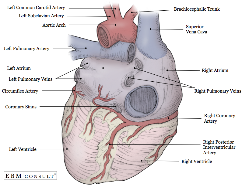
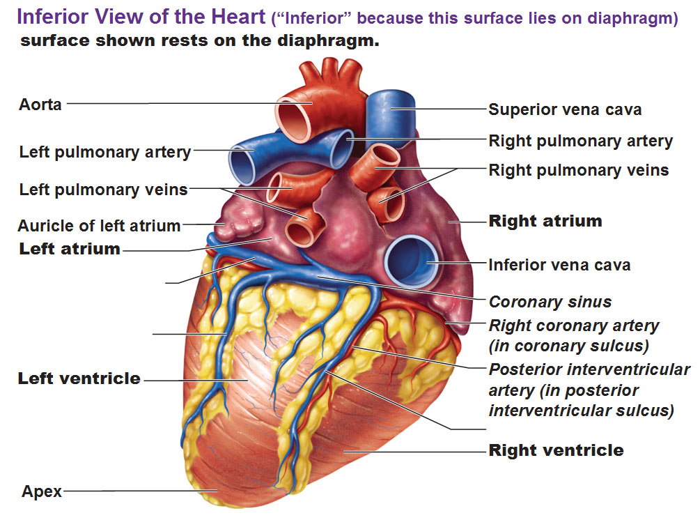
The lungs have a unique blood supply receiving deoxygenated blood from the heart in the pulmonary circulation for the purposes of receiving oxygen and releasing carbon dioxide and a separate supply of oxygenated blood to the tissue of the lungs in the bronchial circulation. How does the heart connect to the lungs. Heart and lung anatomy this image shows the anatomy of the heart and the lungs in relation to each other displaying their different parts and features and the vessels of the heart and their relation to the lungs showing.
Within the mediastinum the heart is separated from the other mediastinal structures by a tough membrane known as the pericardium or pericardial sac and sits in its own space called the pericardial cavity. Lets take a look at some anatomy of the lungs. Anatomy and physiology of heart lung 1.
On the inferior surface the lungs are bordered by the diaphragm. Arch of the aorta 2. Apex the blunt superior end of the lung.
Base the inferior surface of the lung which sits on the diaphragm.
Sample 1 Heart And Lung Diagram Accessible Image Sample Book
 Pulmonary Artery Anatomy Britannica
Heart And Lungs Drawing At Getdrawings Com Free For
 Lung Anatomy Diagram Thorax Lungs Heart Anatomy And
 Cardiovascular System The Heart Anatomy Flashcards Quizlet
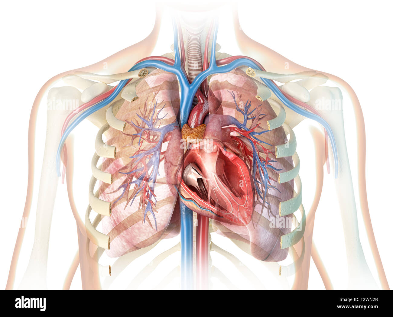 Rib Cage Heart Lungs Stock Photos Rib Cage Heart Lungs
:watermark(/images/watermark_5000_10percent.png,0,0,0):watermark(/images/logo_url.png,-10,-10,0):format(jpeg)/images/atlas_overview_image/38/1IG8yMbrwvj1SsNFasBA_lungs-in-situ_english.jpg) Pulmonary Arteries And Veins Anatomy And Function Kenhub
Pulmonary Arteries And Veins Anatomy And Function Kenhub
 Human Anatomy Medical Poster With Internal Organs Kidney Lung
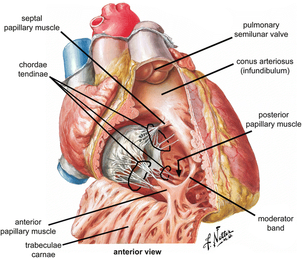 Anatomy Of The Human Heart Springerlink
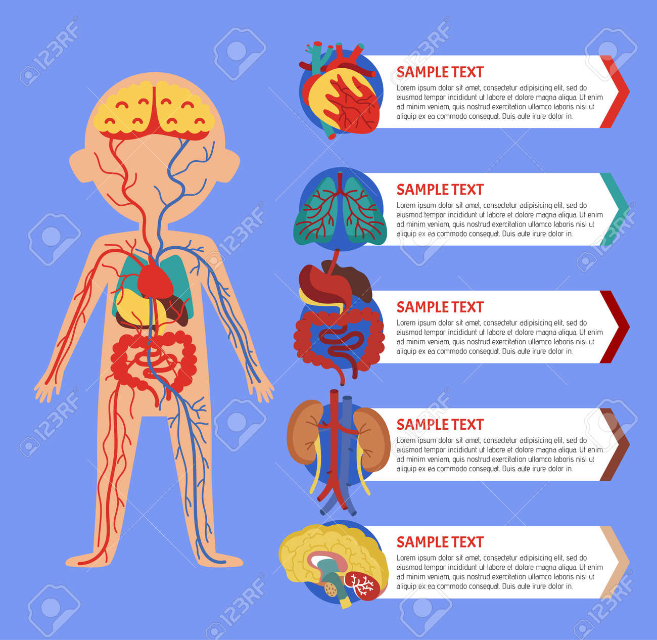 Health Medical Poster With Human Body Anatomy Kidney Lung
1 4 Basic Organs Of The Body Training Manual Hiv I Base
:background_color(FFFFFF):format(jpeg)/images/library/7777/sternocostal-surface-of-the-heart_english.jpg) Pulmonary Arteries And Veins Anatomy And Function Kenhub
Pulmonary Arteries And Veins Anatomy And Function Kenhub
 How The Heart Works Human Heart Diagram Blood Flow To The
 Pulmonary Circulation Wikipedia
 Anatomy Of The Heart And Great Vessels Cardiology Medical
 Human Torso Body Organ Anatomy Model Parts Head Brain Heart Lung Medical Science
 Lungs Heart Posterior View Carlson Stock Art
 The Heart Anatomy Physiology And Function
Heart Lung Transplant Western New York Urology Associates Llc


Belum ada Komentar untuk "Lung Heart Anatomy"
Posting Komentar