L Spine Anatomy
And coccygeal the tail bone. First are the vertebrae of the spine and underneath those are three layers of tough membrane called the meninges.
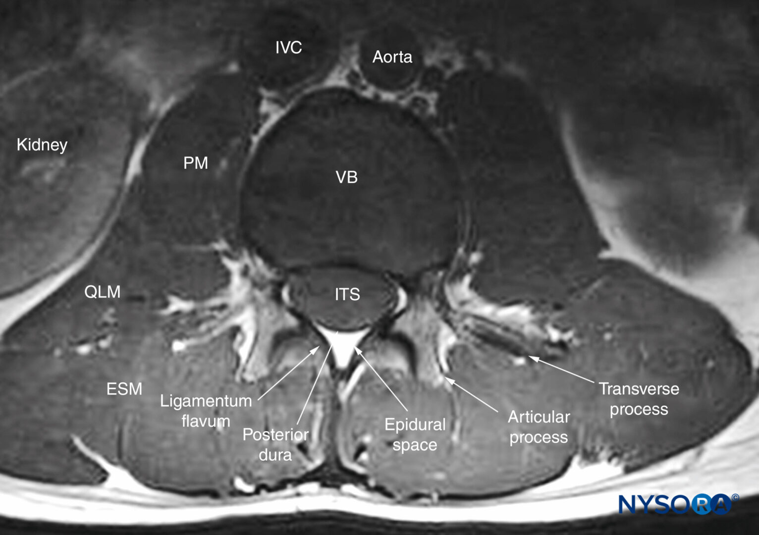
 Lumbar Spine Anatomy On Mri Magnetic Resonance Imaging
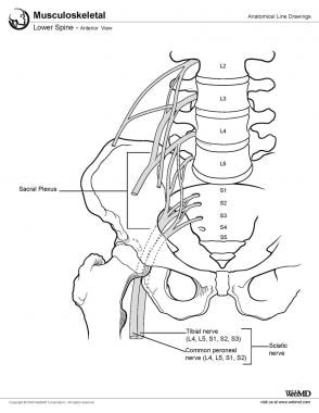
Sacral attached to the pelvis.
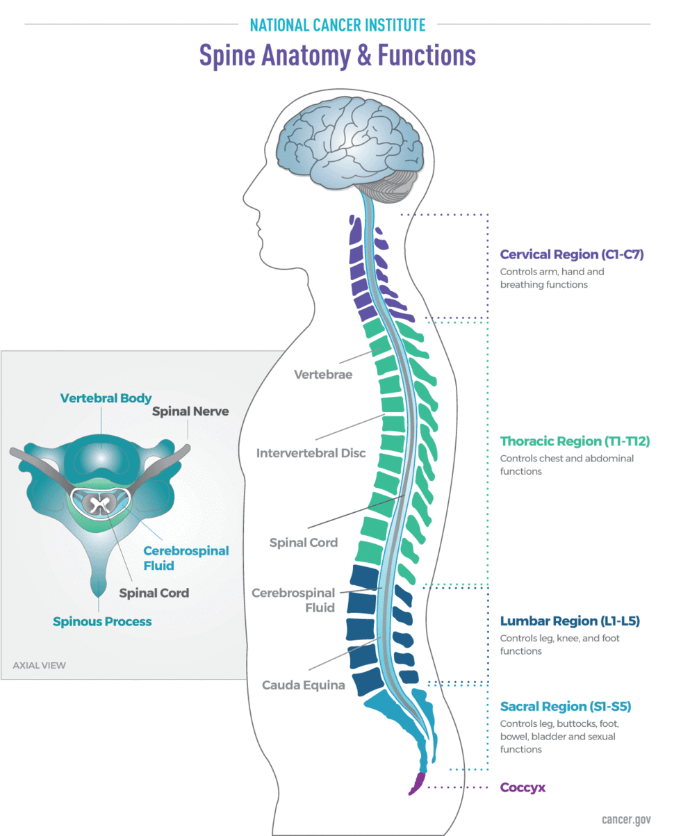
L spine anatomy. Each region has a number of vertebral bones. When healthcare professionals refer to the spine theyre generally talking about the 24 vertebrae that form an elegant double s shaped line. This section of the website will explain large and minute details of lumbar spine sagittal cross sectional anatomy.
The vertebral column also known as the backbone or spine is part of the axial skeleton. Definition articulation refers to the motion that occurs between joints. The spinal cord like the brain has two major layers of protection.
There are hundreds of individual ligaments in the spine. Thoracic in the chest. Ligaments typically joint bones to other bones to stabilize them to one another.
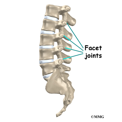
The spine is an intricate set of bones muscles nerves and discs. Understanding the anatomy of the spine is a fundamental basis to knowing your spinal condition. For example certain facet joints in the spine allow for up down side to side and twisting movements.
This mri lumbar spine cross sectional anatomy tool is absolutely free to use. The five vertebrae of the lumbar spine l1 l5 are the biggest unfused vertebrae in the spinal column enabling them to support the weight of the entire torso. As mentioned previously the anatomy of the spine is quite complex.
Thomas schuler spine surgeon chief executive officer and founder of virginia spine institute has provided two videos below providing an overview of spinal anatomy. The vertebral column is the defining characteristic of a vertebrate in which the notochord a flexible rod of uniform composition found in all chordates has been replaced by a segmented series of bone. The lumbar spines lowest two spinal segments l4 l5 and l5 s1 which include the vertebrae and discs bear the most weight and are therefore the most prone to degradation and injury.
The spine also has joints that are similar to knees elbows and other joints. Lumbar spine joints. It is divided into five regions.
Vertebrae separated by intervertebral discs. In the spine tendons connect muscles to the vertebrae. The spine also known as the vertebral column or spinal column is a column of 26 bones in an adult body 24 separate vertebrae interspaced with cartilage and then additionally the sacrum and coccyx.
Spinal anatomy the regions of the spine. Lumbar spine anatomy and pain. Ligaments are strong tough bands that are typically not very flexible.
The ligaments and tendons help to stabilize the spine and guard against excessive movement in any one direction.
 Learn All About Lumbar Spine Anatomy From A World Renowned
 Spinal Sonography And Applications Of Ultrasound For Central
 Lumbar Spine Anatomy Overview Gross Anatomy Natural Variants
 Lumbar Spine Anatomy Eorthopod Com
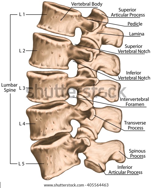 Lumbar Spine Structure Vertebral Bones Lumbar Science
 Read Your Mri Basic Education From A World Renowned Spine
Taconic Spine Anatomy Library Spine Center For Back Pain
 Lumbar Spine Lower Back Anatomy Function Problems
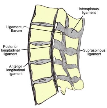 Lumbar Spine Anatomy Overview Gross Anatomy Natural Variants
 Brain And Spine Tumor Anatomy And Functions National
 Lumbar Spine Anatomy Spine Orthobullets
 Learn All About Lumbar Spine Anatomy From A World Renowned
 Lumbar Spine Anatomy Posterior View Anatomy Craniosacral
 Anatomy Of The Spine Teachpe Com
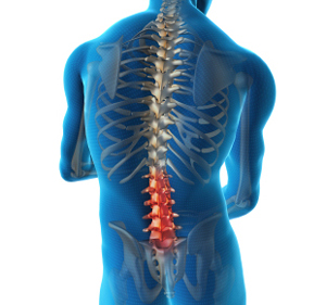 Lumbar Spine Anatomy Washington Back Bone Pain Treatment
 Radiological Anatomy Of The Lumbar Spine X Ray Mri Ct Covered
 Mri Spine Anatomy Free Mri Axial Cervical Spine Anatomy
 Lumbar Spine Anatomy On Mri Magnetic Resonance Imaging
Lumbar Spine Radiographic Anatomy Wikiradiography
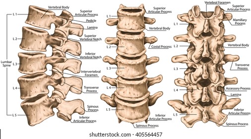 Lumbar Vertebra Images Stock Photos Vectors Shutterstock


Belum ada Komentar untuk "L Spine Anatomy"
Posting Komentar