Nerves Of The Leg Anatomy
There are two main nerves in the leg. The tibial nerve passes into the foot running posterior to the medial malleolus and beneath the flexor retinaculum.
Major nerves of the hip thigh lower leg and foot.
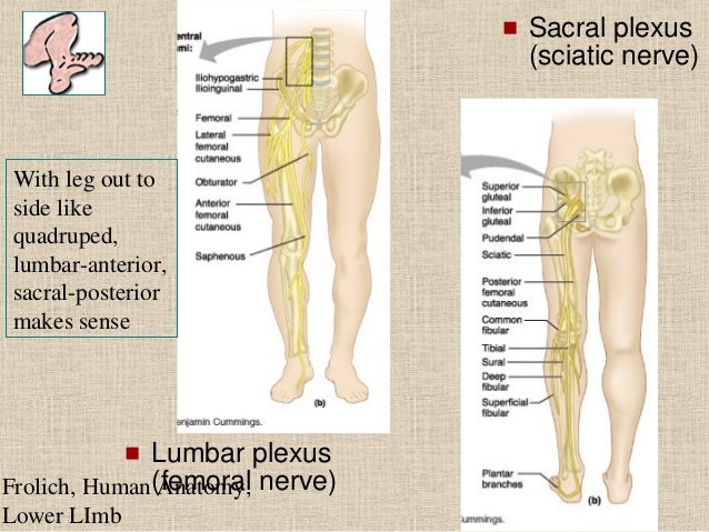
Nerves of the leg anatomy. Atlas main nerves of the lower extremity. Teachme anatomy part of the teachme series the medical information on this site is provided as an information resource only and is not to be used or relied on for any diagnostic or treatment purposes. The superior l4 s1 and inferior gluteal nerves l5 s2 innervate the gluteus muscles and the tensor fasciae latae.
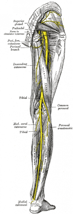
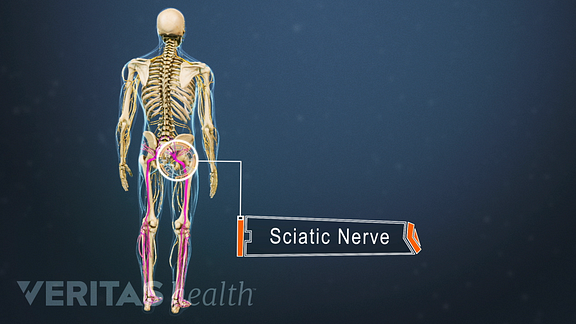
Major nerves of the hip thigh lower leg and foot. In this article you will find the anatomy branches and mnemonics related to the lumbar plexus. Along its route through the legs the sciatic nerve splits into the tibial and common fibular peroneal nerves which in turn split into many smaller nerves in the legs and feet.
Neurovasculature of leg and knee watch video. Sensory nerves are of course present throughout the lower extremities. Anatomy course function and clinical significance of the sciatic nerve.
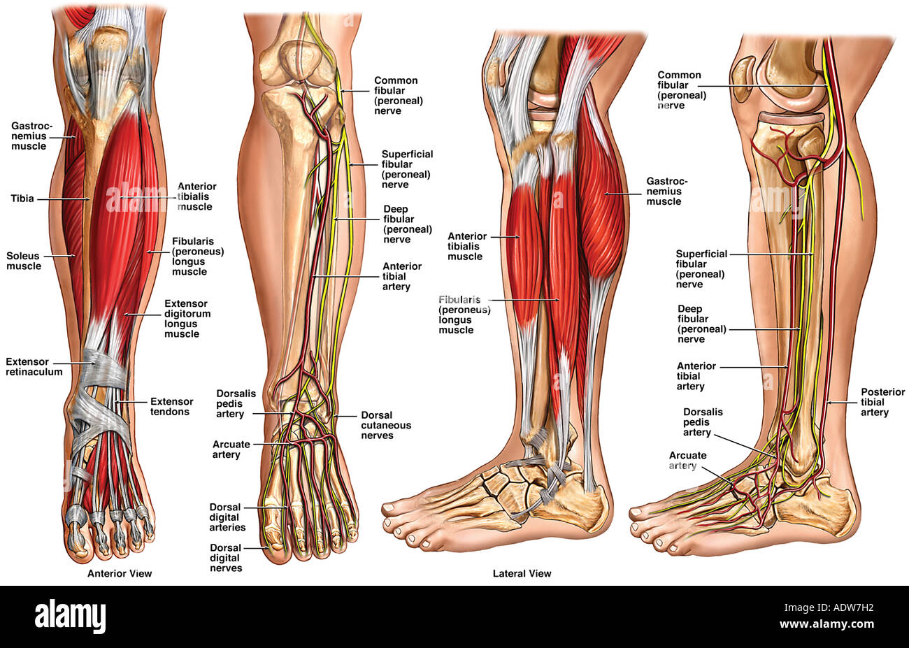
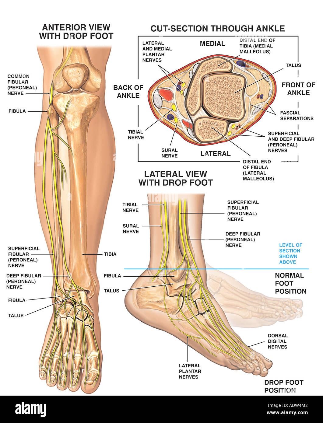
In terms of innervation the leg receives it via the common fibularperoneal tibial and saphenous nerves. The nerves of the leg and foot serve to propel the body through the actions of the legs feet and toes while maintaining balance both while the body is moving and when it is at rest. However with the exception of the bottom of the foot they play a lesser role here than in the upper extremities.
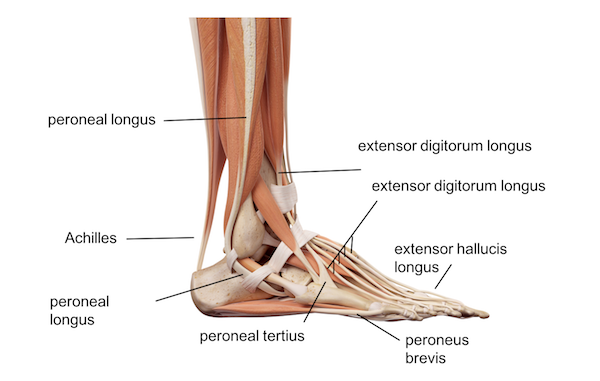
Lateral and medial gastrocnemius. The location of the nerve pain can determine which of the nerves is injured. Branches of the tibial branch of the sciatic nerve stimulate muscles in the lower leg including the.
The nerves of the leg can have many nerve roots and when pain or discomfort is felt in these areas it usually indicates a compressed or pinched nerve. The femoral nerve serves the front and the sciatic nerve controls the back of the leg. The first two are branches of the sciatic nerve while the latter stems from the femoral nerve.
The nerves of the leg and foot. These three nerves divide further to supply the various structures of the leg. At this level the posterior tibial nerve divides into medial and.
The nerves of the foot help move the body and keep balance both while its moving and at rest. The nerves of the sacral plexus pass behind the hip joint to innervate the posterior part of the thigh most of the lower leg and the foot.
 Cutaneous Nerve Blocks Of The Lower Extremity Nysora
 Venous Drainage And Cutaneous Innervation Of The Lower Limb
 Image Tibial Nerve For Term Side Of Card Muscle Anatomy
 Anatomy Of The Lower Leg Stock Photo 7712337 Alamy
 Femoral Nerve Block Landmarks And Nerve Stimulator
 5 Anatomy Of The Obturator Nerve In The Leg Image By
 Posterior Anatomy Right Leg Medical Art Works
 Chapter 37 Leg The Big Picture Gross Anatomy
Fg Anatomy G50 Nerve Lesions Of The Lower Extremity Anatomy
 Sciatic Nerve Nerve Anatomy Nerves In Human Body Sciatic
 Common Peroneal Nerve Physiopedia
 Leg Nerves Stock Photos Leg Nerves Stock Images Alamy
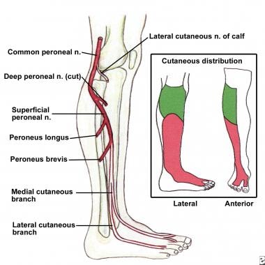 What Is The Anatomy Of The Lower Leg Affected By Foot Drop
 Common Peroneal Nerve Neurologyneeds Com
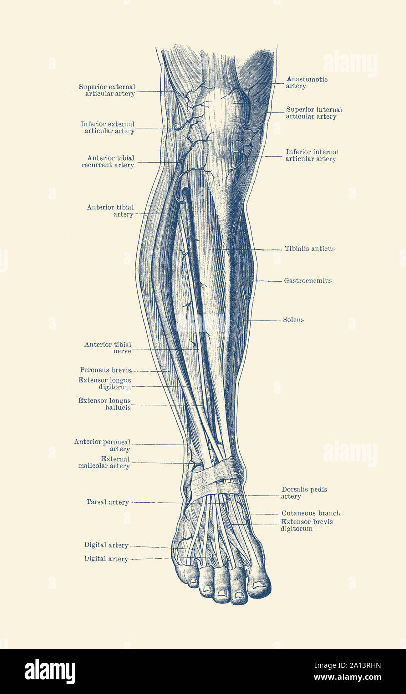 Human Leg Nerves Stock Photos Human Leg Nerves Stock
 Core Anatomy Winding Round To Foot Drop Which Nerve Is
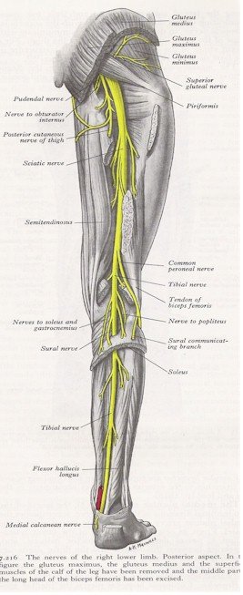 Nerves Of The Leg Posterior View Myfootshop Com
 Leg Muscles Anatomy With Nerves
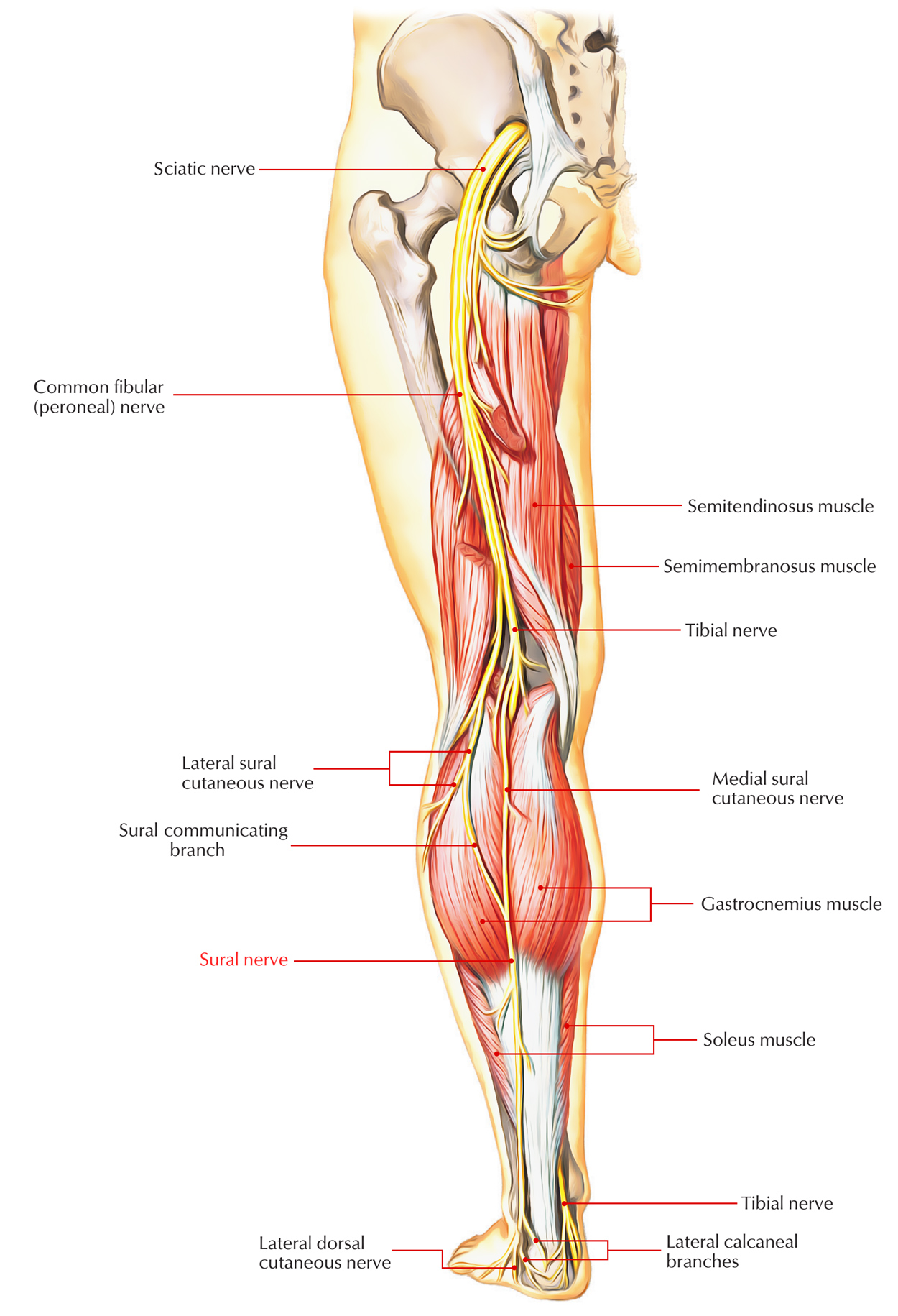 Easy Notes On Sural Nerve Learn In Just 4 Minutes
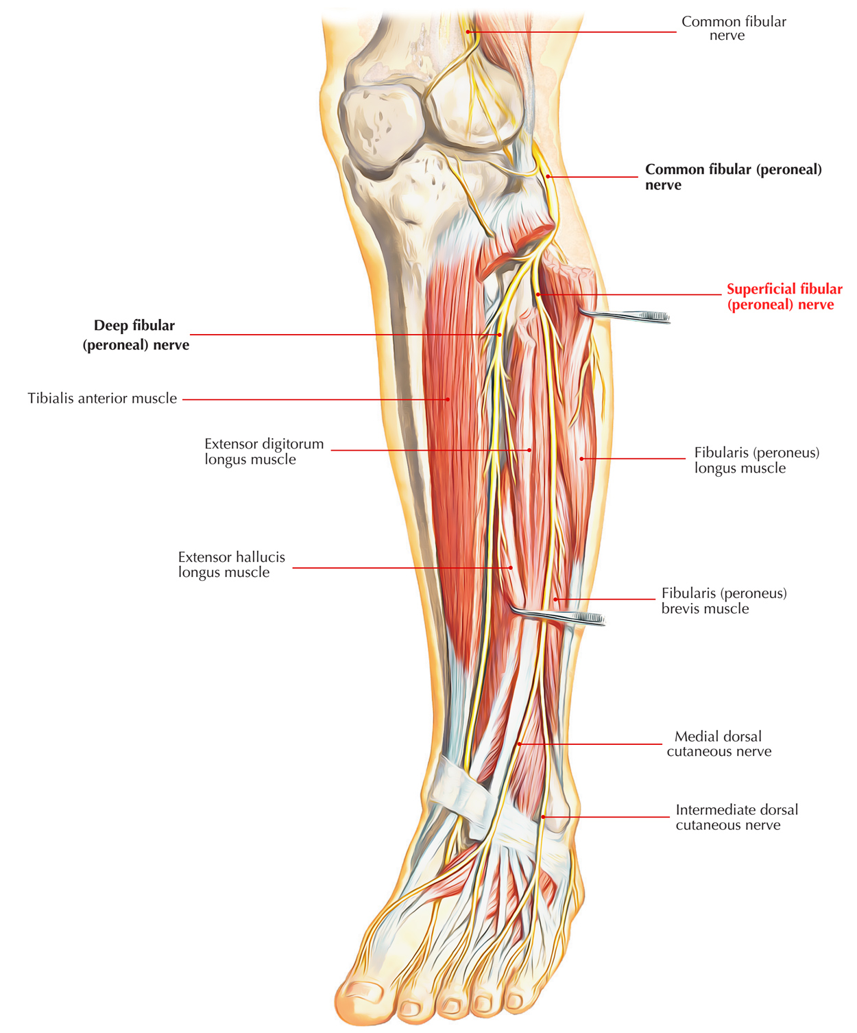 Easy Notes On Superficial Peroneal Nerve Learn In Just 3
:background_color(FFFFFF):format(jpeg)/images/library/11085/_990_LEx-cropped.png) Lower Extremity Anatomy Bones Muscles Nerves Vessels
Lower Extremity Anatomy Bones Muscles Nerves Vessels
 Nerves Of The Legs From Foundations Of Anatomy Physiol
 Nerves Of The Leg And Foot Interactive Anatomy Guide
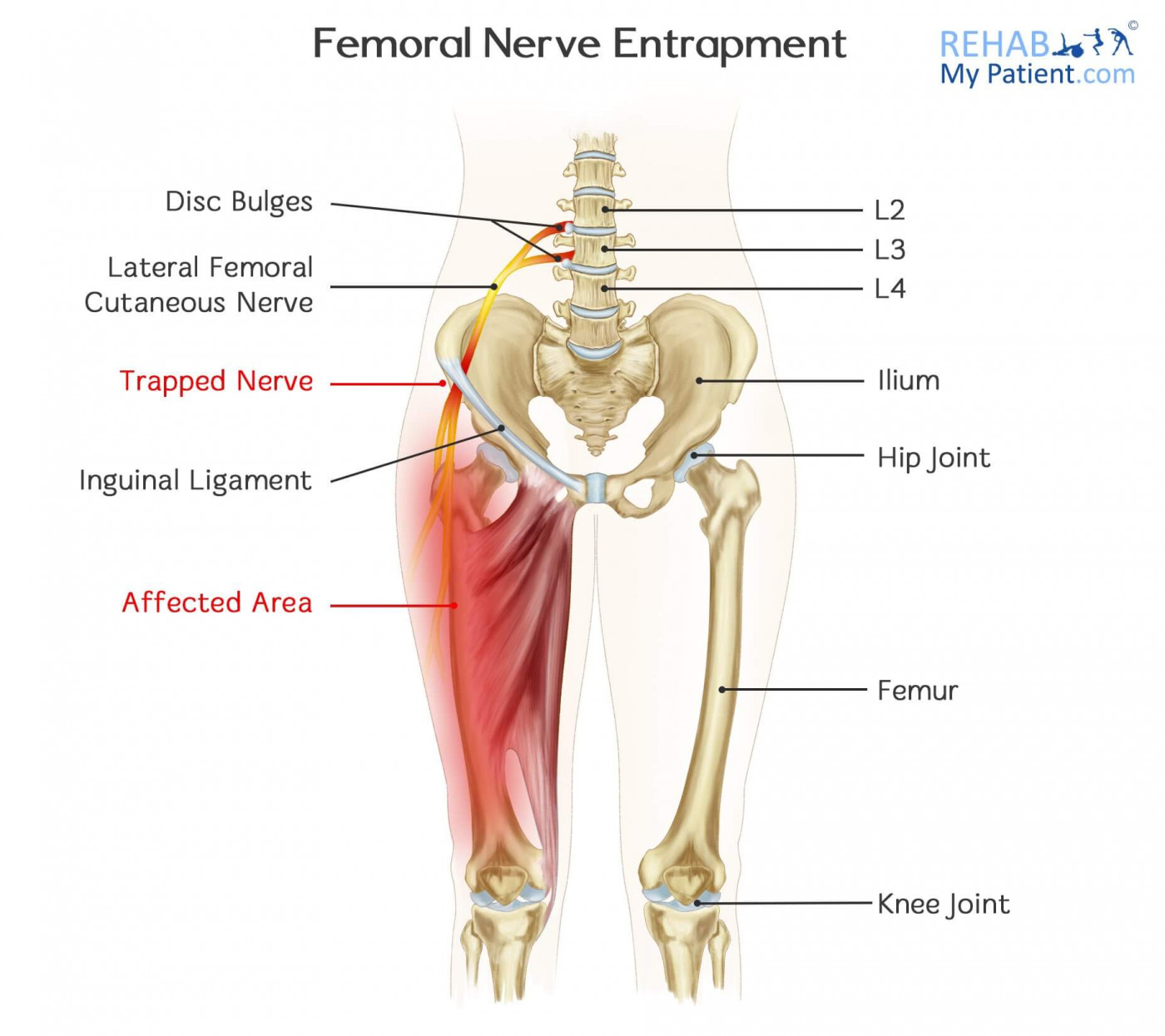 Femoral Nerve Entrapment Rehab My Patient


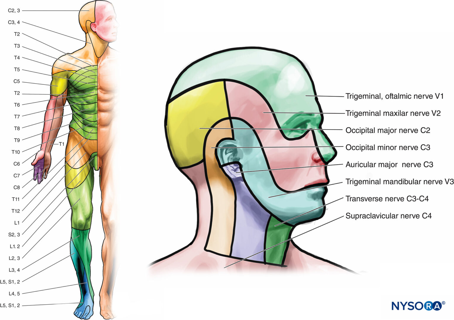 Functional Regional Anesthesia Anatomy Nysora
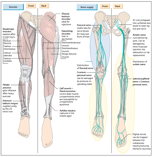 Anatomy Of The Leg Musculoskeletal Key
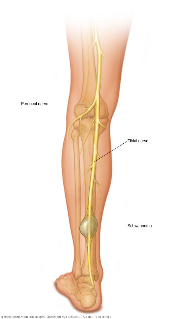 Peripheral Nerve Tumors Symptoms And Causes Mayo Clinic

Belum ada Komentar untuk "Nerves Of The Leg Anatomy"
Posting Komentar