Distal Radius Anatomy
The ulna bone may also be broken. Distal radius fractures broken wrist the radius is the larger of the two bones of the forearm.
 Distal Radius Fracture
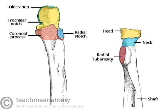
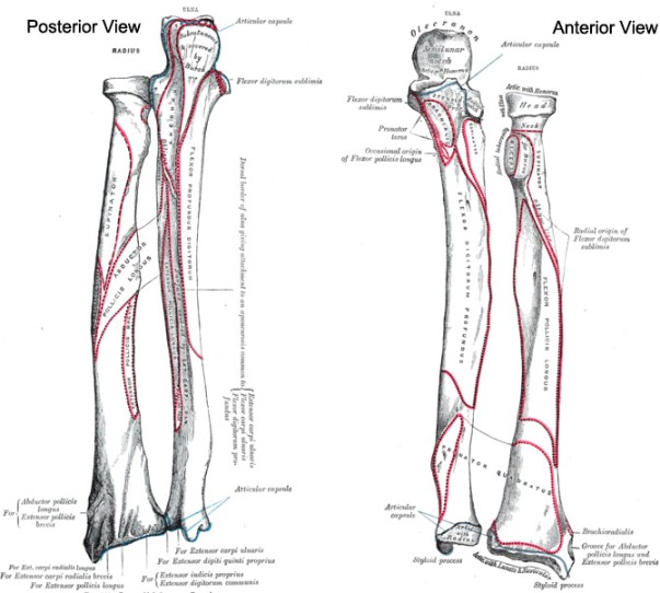
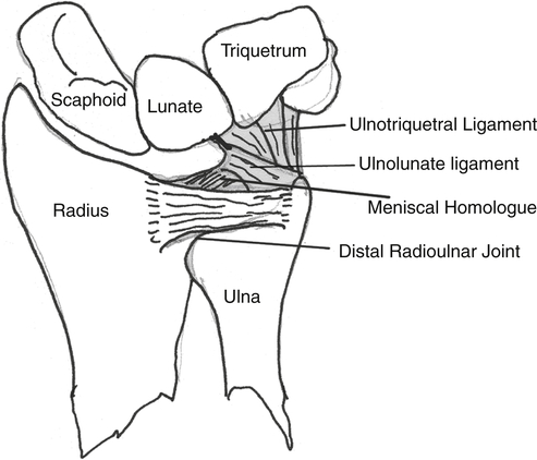
The distal radius rotates about the ulnar head by means of the sigmoid notch which is concave with well defined dorsal palmar and distal margins.

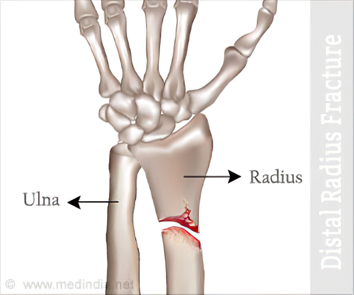
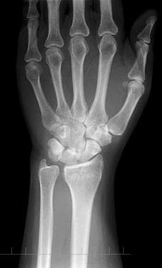
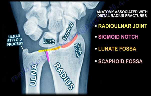
Distal radius anatomy. X rays are the most common method in evaluating a fracture if the bone has been displaced. When the radius breaks near the wrist it is called a distal radius fracture. The medial surface of the distal radius consists of the ulnar notch and the articular surface for the ulnar head figure 1.
Distal radius fractures are very common. In the distal region the radial shaft expands to form a rectangular end. A ct scan.
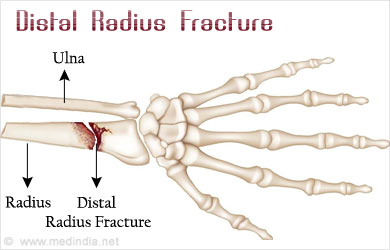
The radius is one of two forearm bones and is located on the thumb side. The part of the radius connected to the wrist joint is called the distal radius. Symptoms include pain bruising and rapid onset swelling.
A fracture of the distal radius occurs when the area of the radius near the wrist breaks. Radiographs of the affected wrist are shown in figure a. Distal region of the radius.
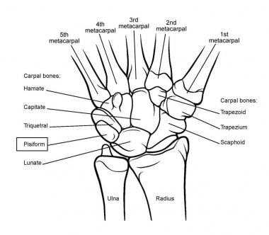
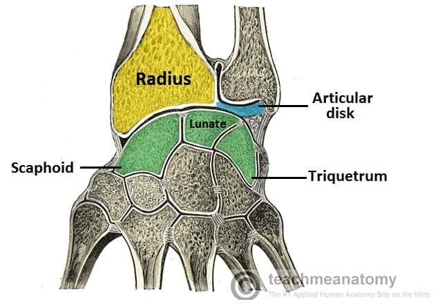
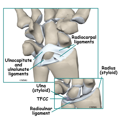
The break usually happens due to falling on an outstretched or flexed hand. A thorough physical examination is important in identifying any noticeable deformities swelling. In the medial surface there is a concavity called the ulnar notch which articulates with the head of ulna forming the distal radioulnar joint.
Distal radius fractures can be reset either with surgery open reduction or without it closed reduction. General features of distal radius anatomy. In younger people these fractures typically occur during sports or a motor vehicle collision.
Radial head dislocation is another common injury associated with the bone 10. A colles fracture or distal radius fracture is often called a broken wrist technically its a break in the larger of the two bones in your forearm. The bone breaks on the lower end.
This article discusses the recovery process for both approaches plus the pain management tactics that can be used for all patients. The end toward the wrist is called the distal end. After soft tissue swelling subsides open reduction and internal fixation of the distal radius is performed.
Obq1378 a 45 year old construction worker sustains a fall and presents with an isolated injury to his upper extremity. In fact the radius is the most commonly broken bone in the arm. The wrist may be deformed.
A distal radius fracture also known as wrist fracture is a break of the part of the radius bone which is close to the wrist. The bone may also be affected by arthritis of the wrist or elbow joints. The radius is considered the most commonly fractured bone in the human body with distal radius fractures being the most common form of radial fracture 9.
Diagnostic methods that a physician may use to help diagnose a distal radius fracture include. The lateral side projects distally as the styloid process.
 4 Ways To Classify Distal Radius Fractures Wikihow
![]() Radius And Ulna Anatomy And Clinical Notes Kenhub
Radius And Ulna Anatomy And Clinical Notes Kenhub

 Distal Radius Fracture Types Symptoms Treatment
 The Radioulnar Joints Teachmeanatomy
 Distal Radius Fractures Trauma Orthobullets
 Wrist Joint Anatomy Overview Gross Anatomy Natural Variants
 The Radius Proximal Distal Shaft Teachmeanatomy
 Distal Radius Fracture

 Distal Radius Fracture Types Symptoms Treatment
 X Wrist Startradiology
 Distal Radius Fracture
 Bones Of The Upper Limb Anatomy And Physiology I
 Figure 3 From Surgical Approaches To The Distal Radius
 Distal Radius Fracture
![]() Radius And Ulna Anatomy And Clinical Notes Kenhub
Radius And Ulna Anatomy And Clinical Notes Kenhub
 Bones Of The Upper Limb Anatomy And Physiology
 Distal Radial Fracture Imaging Practice Essentials
 X Wrist Startradiology
 Distal Forearm Approach Volar Anterior Approach M
 Distal Radius Fracture Wikipedia
 Distal Radial Fracture Colles Fracture Radiology Case
Distal Ulna An Overview Sciencedirect Topics
 Radius Anatomy Pictures And Information
 Distal Radius Fractures Reconstruction Approaches Planning
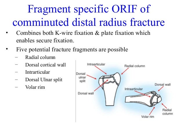
 Comminuted Intraarticular Distal Radius Fractures When To Fix Span Or Close Reduce
 Distal Forearm Decision Support Ao Surgery Reference

 Radius Bone Anatomy Bone And Spine
 Wrist Fractures Musculoskeletal Key
 Common Types Of Distal Radius Fractures Nabil Ebraheim
![]() Radius And Ulna Anatomy And Clinical Notes Kenhub
Radius And Ulna Anatomy And Clinical Notes Kenhub
Belum ada Komentar untuk "Distal Radius Anatomy"
Posting Komentar