Fetal Ultrasound Anatomy
By the 20th week of pregnancy the baby can weigh up to 11 ounces and measure 10 inches outstretched. Although real time scanning of the gravid uterus quickly allows the observer to determine fetal lie and presentation this maneuver of identifying specific right and left sided structures within the fetal body forces one to determine fetal position accurately and identify normal and pathologic fetal anatomy.
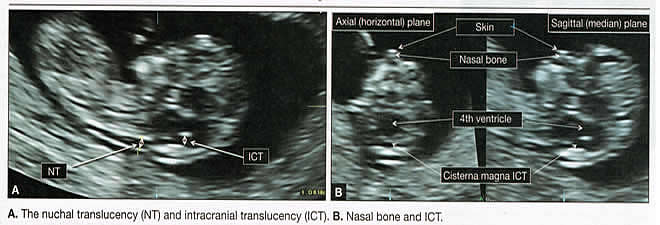
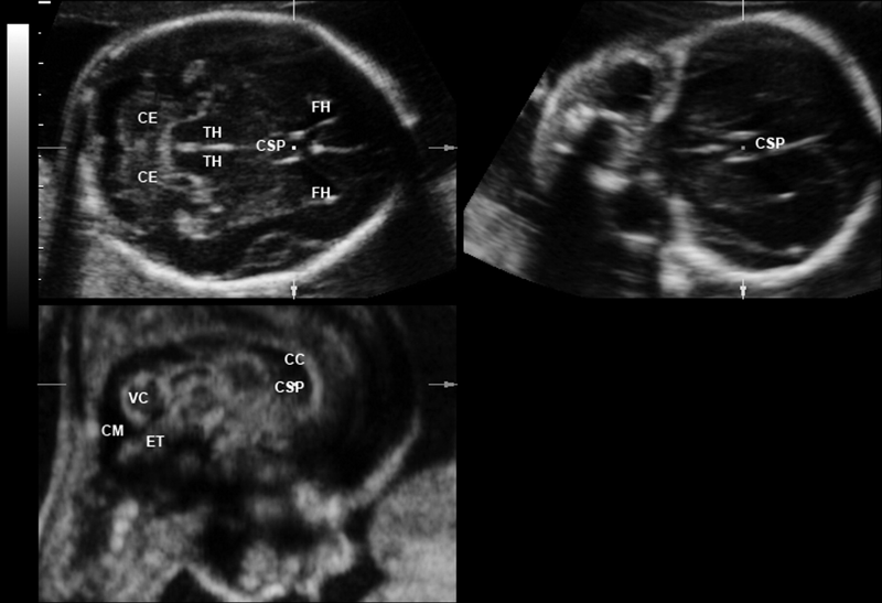
 Normal First Trimester Anatomic Features Of The Fetal Brain
Outline superficial anatomy of the fetus 161 musculoskeletal system 161 cardiovascular system 183 gastrointestinal system 191 respiratory system 197 genitourinary system 199 central nervous system 200 summary of key points continued advancement of ultrasound technology including increase in frequency and choice of focal position have improved visualization of fetal anatomy and.

Fetal ultrasound anatomy. Ultrasound of the foetal heart showing scanning technique protocols chambers vies outflow tracts and normal fetal heart anatomy. The second trimester scan is a routine ultrasound examination in many countries that is primarily used to assess fetal anatomy and detect the presence of any fetal anomalies. This sonogram is used to determine fetal anomalies the babys size and weight and also to measure growth to ensure that the fetus is developing properly.
The anatomy scan is a level 2 ultrasound which is typically performed between 18 and 22 weeks. The second trimester extends from 13 weeks and 0 days to 27 weeks and 6 days of gestation although the majority of these studies are performed between 18 and 23 weeks. When the pregnancy hits the 20th week of gestation an anatomy ultrasound is often ordered.
Determining fetal sex the gender of your babybabies can usually be determined at this ultrasound. Fetal ultrasound images can help your health care provider evaluate your babys growth and development and monitor your pregnancy. In some cases the baby may have their legs crossed or be facing away from the abdomen and thus the sexual organs will not be visible during the anatomic ultrasound.
A fetal ultrasound sonogram is an imaging technique that uses sound waves to produce images of a fetus in the uterus. In some cases fetal ultrasound is used to evaluate possible problems or help confirm a diagnosis. Other than finding out the sex of your baby if you want to know the ultrasound technician will be.
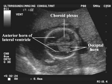
 Fetal Lateral Ventricle Measurements How To Measure Posterior Ventricle For Ventriculomegaly
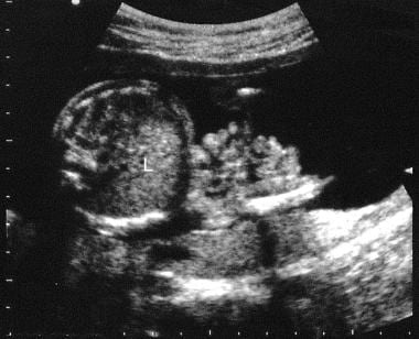
 Second Trimester Fetal Development Images Of Your Growing
:max_bytes(150000):strip_icc()/10coupland18usboy-56a76b945f9b58b7d0ea5792-597f670d685fbe001165639e.jpg) Ultrasound Images Of What Your Growing Boy Looks Like
Ultrasound Images Of What Your Growing Boy Looks Like
 Gastroschisis Imaging Overview Radiography Computed
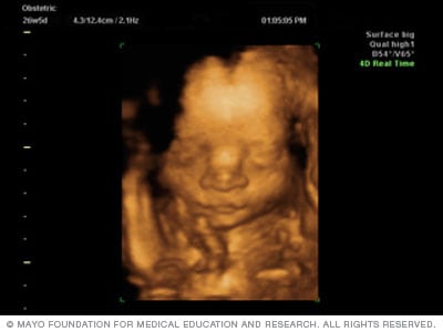 Fetal Ultrasound Mayo Clinic
 Ultrasound And Color Doppler Videos Normal Third Trimester
 Ultrasound Atlas Glowm
 Atlas Of Fetal Sectional Anatomy With Ultrasound And

Fetal Anomalies Associated With Breech Presentation Fetal
 Ultrasound Level One Services Heavenly 3d 4d Ultrasound
 Image Iq Anatomy Of The Fetal Spine Obgyn Net
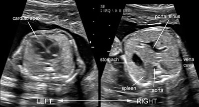
 Sectional Fetal Anatomy In Ultrasound

 Fetal Heart Ultrasound How To
 Why More Women Should Decline Their First Trimester
 The Radiology Assistant Neonatal Brain Us
:max_bytes(150000):strip_icc()/992aaaaaap-56a767755f9b58b7d0ea295f.jpg) Ultrasound Images Of What Your Growing Boy Looks Like
Ultrasound Images Of What Your Growing Boy Looks Like
 Fetal Anatomy Screening 1 Sono Care Of East Texas Tyler
 Ultrasound Evaluation Of Normal Fetal Anatomy Radiology Key
 The Radiology Assistant Normal Values Ultrasound
 Why More Women Should Decline Their First Trimester
 A Gallery Of High Resolution Ultrasound Color Doppler 3d
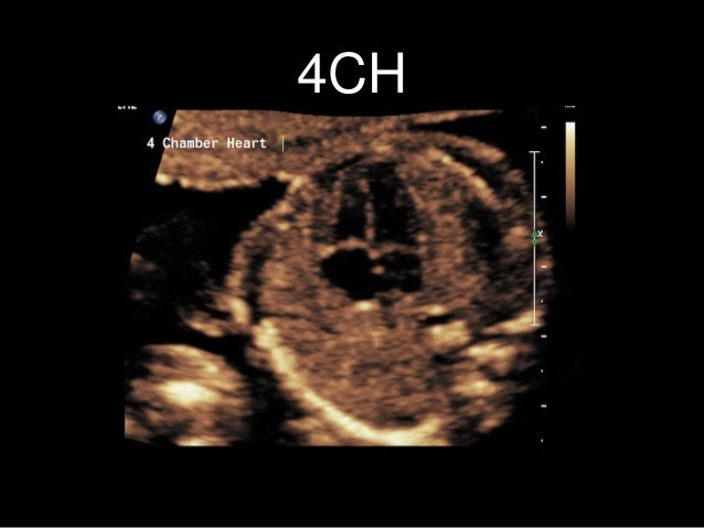 Labelled Fetal Heart Ultrasound

Belum ada Komentar untuk "Fetal Ultrasound Anatomy"
Posting Komentar