Lateral Chest X Ray Anatomy
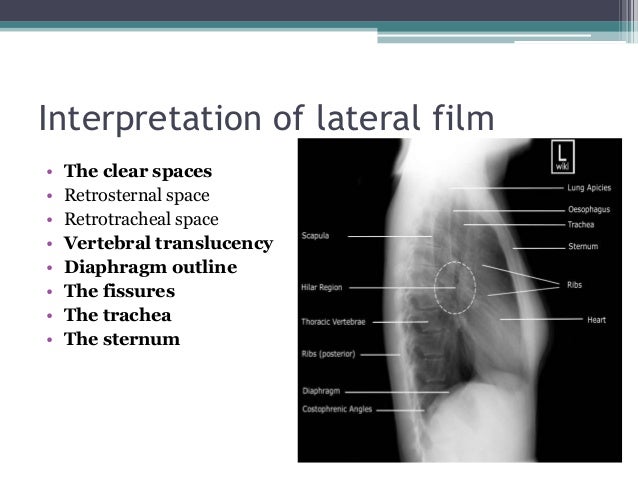
The lateral chest view examines the lungs bony thoracic cavity mediastinum and great vessels. Lobes and fissures this cut out of a lateral chest x ray shows the positions of the lobes of the right lung roll over the image.
:watermark(/images/watermark_5000_10percent.png,0,0,0):watermark(/images/logo_url.png,-10,-10,0):format(jpeg)/images/atlas_overview_image/804/3NCDt3BMfHeJjE6EgeXrw_chest-x-ray-pa-view_english.jpg) Normal Chest X Ray Anatomy Tutorial Kenhub
Normal Chest X Ray Anatomy Tutorial Kenhub
The lateral chest view may be performed as an adjunct to a frontal chest radiograph in cases where there is diagnostic uncertainty.

Lateral chest x ray anatomy. As part of the standard two view chest examination patients usually have an upright frontal chest radiograph and an upright left lateral view of the chest. Identifying the exact lobe of a lobar pneumonia in the right lung. On the left the oblique fissure is in a similar position but there is usually no horizontal fissure and so there are only two lobes on the left.
The video will describe anatomy on a lateral view of chest. A left lateral chest x ray the patients left side is against the film is of great diagnostic value but is sometimes ignored by beginners because of their lack of familiarity with the findings visible in that projection. Often a lateral view usually accompanies a paap chest x ray.
This can be helpful in settings where the single view is limited in localizing pathology ie. This allows the x ray beams to travel from the emitter through the patient from right to left to the receptor on the other side. To take left lateral films the patient stands or sits upright with his or her arms raised and turns 90 degrees so that the left side faces the receptor.
Please see disclaimer on my website.
What To Look For On A Chest X Ray Slideshow
 Pin By So Heavenly On X Ray Radiology Student Radiology
 Basics Of Chest X Ray Brian S Radiology Learning Diary

 Approach To The Chest X Ray Cxr Undergraduate Diagnostic
Thoracic Spine Radiographic Anatomy Wikiradiography
 The Radiology Assistant Chest X Ray Basic Interpretation
 Radiography Makes Use Of High Energy Photons Called X Rays
Image Of Sarcoidosis Lateral Chest Radiograph
Interpreting A Chest X Ray Stepwards
 Method To Read Lateral Chest X Rays Undergroundmed
 Chest Radiography Lecture 2 Ppt Video Online Download
 Chest X Ray Anatomy Diaphragm Hemidiaphragms Lateral
 Normal Chest X Ray Litfl Medical Blog Labelled Radiology
 Interpreting Chest X Rays Ct Scans And Mris Respiratory
Interpreting A Chest X Ray Stepwards
 Basics Of Chest X Ray Brian S Radiology Learning Diary
Chest Radiographic Anatomy Wikiradiography
 Normal Chest X Ray Lobes Illustration Radiology Case
 Cardiomediastinal Anatomy On Chest Radiography Annotated
Lateral Chest Paravertebral Gutter Positioning Technique
 Normal Chest Pa And Lat Radiographic Views Chest X Ray A


Belum ada Komentar untuk "Lateral Chest X Ray Anatomy"
Posting Komentar