Pelvic Xray Anatomy
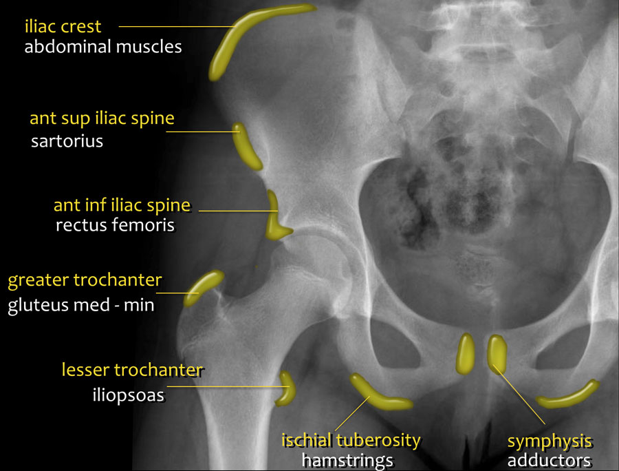
The ap pelvis view is part of a pelvic series examining the iliac crest sacrum proximal femur pubis ischium and the great pelvic ring. Asis anterior superior iliac spine attachment site for sartorius muscle.
 Radiographs Of The Dog
Mands thorough break down of this commonly used ed diagnostic the pelvic xr.

Pelvic xray anatomy. An x ray of the pelvis focuses specifically on the area between your hips that holds many of your reproductive and digestive organs. Ct of the pelvis is the technique of choice for evaluating complex fracture patterns degree of displacement and soft tissue injury. The sacroiliac joints should be symmetrical joint space range 2 4 mm.
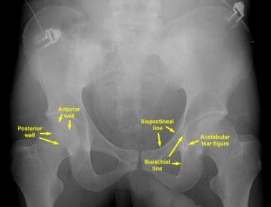
Ilium ischium and pubis connected by the triradiate cartilage. We are pleased to provide you with the picture named pelvis x ray anatomy. Each hemi pelvis bone comprises 3 bones the ilium white pubis orange and ischium blue the 3 bones fuse to form the acetabulum the pelvic portion of the hip joint.
It is of considerable importance in the management of severely injured patients presenting to emergency departments 1. Pelvic xrays are a key component of trauma fractures and dislocations seen every day in the ed but when is the last time you went back over the anatomy and radiographic tips and tricks of the pelvic radiograph. Its primary function is the transmission of forces from the axial skeleton to the lower limbs as well as supporting the pelvic viscera.
If either joint space is widened think main pelvic ring fracture. The series is used most in emergency departments during the evaluation of multi trauma patients due to the complex anatomy the ap projection covers. Your pelvis is made up of three bones the ilium ischium and.
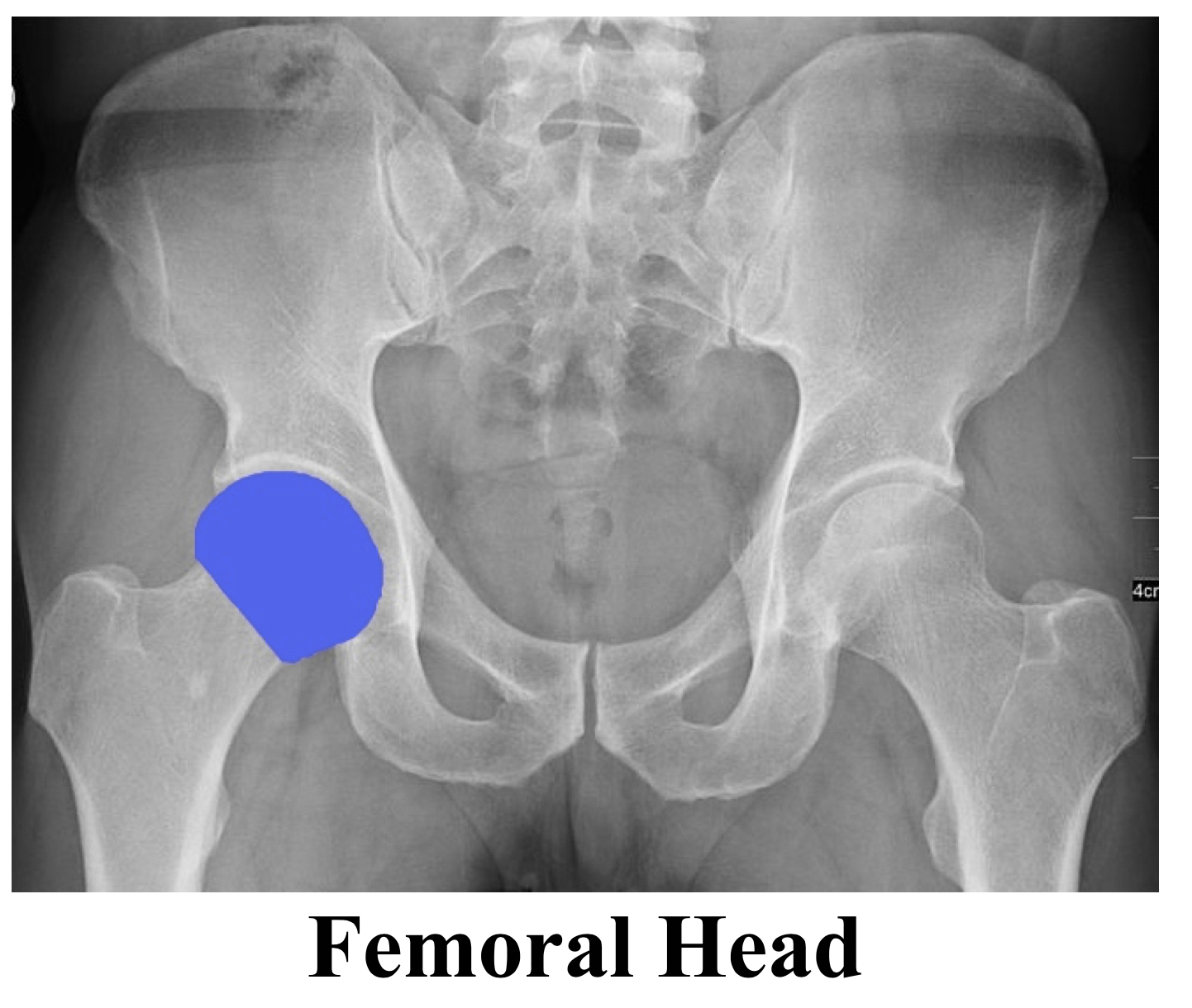
Pelvis x ray anatomy in this image you will find the sacroiliac joint acetabular obturator foramina greater trochanter pubic symphysis femoral heads lesser trochanters in it. Until puberty each hip bone consists of three separate bones yet to be fused. Hemi pelvis anatomy normal ap.
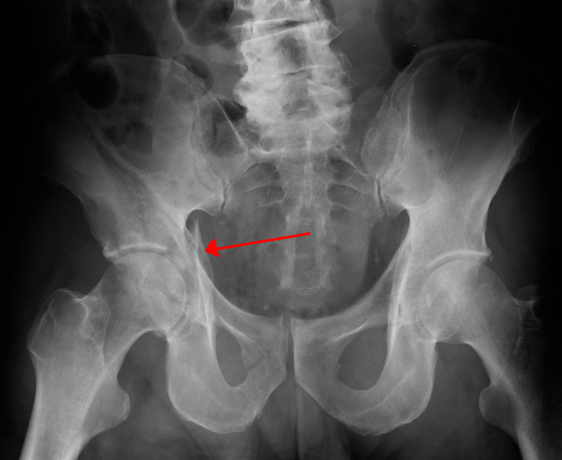
The pelvis series examines the main pelvic ring obturator foramina sacroiliac joints symphysis pubis acetabulum sacral foramina and the proximal femur. Symptoms from fractures of the hip acetabulum and pelvis may be quite similar thus a full ap pelvis radiograph including the hip must be obtained if any of the above fractures are expected. The symphysis pubis joint space should be 5 mm.
 Sacroiliac Luxation In Dogs And Cats Minimally Invasive
 Radiographic Anatomy Of Adult Pelvis Orthopaedicsone
 Radiologic Evaluation Of The Pelvis And Hip Fundamentals

Ao Surgery Reference
 Presentation1 Pptx Radiological Anatomy Of The Abdomen And
 Pelvis Series Radiology Reference Article Radiopaedia Org
 Pelvis Radiograph Right Judet View Anatomy Quiz
 Back To Basics Pelvic Xrays Taming The Sru
 Skeletal Trauma
 How To Read Pelvic X Rays International Emergency Medicine
 What Is The Anatomy Of The Pelvis Relevant To Acetabulum
 Pelvic Ring Fractures Trauma Orthobullets
 Back To Basics Pelvic Xrays Taming The Sru
 Acetabular Fracture Wikipedia
 Radiographs Of The Dog
 Back To Basics Pelvic Xrays Taming The Sru

 Dislocated Hip Symptoms Diagnosis And Treatments Hss

 The Pelvis And Hip
 Human S Pelvis And Hip Joints Stock Photo Image Of Anatomy
 The Radiology Assistant Hip Pathology In Children
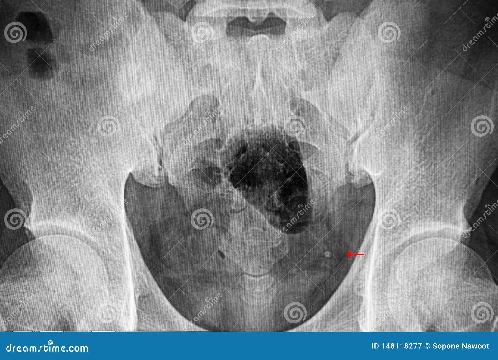 Pelvic Xray Film Showing Distal Ureterc Stone Stock Image

 Pelvic X Ray Normal Different Ages Radiology Case
 Adult Normal Pelvis Acetabular Radiological Anatomy
Belum ada Komentar untuk "Pelvic Xray Anatomy"
Posting Komentar