Cervical Spine Xray Anatomy
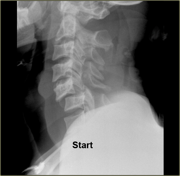
Right anterior oblique view. The interposition grafts are.
 Cervical Myelopathy Spine Orthobullets
Line 1 extends from the top of the anterior cortex of the peg to the anterior margin of the body of t1 vertebra.

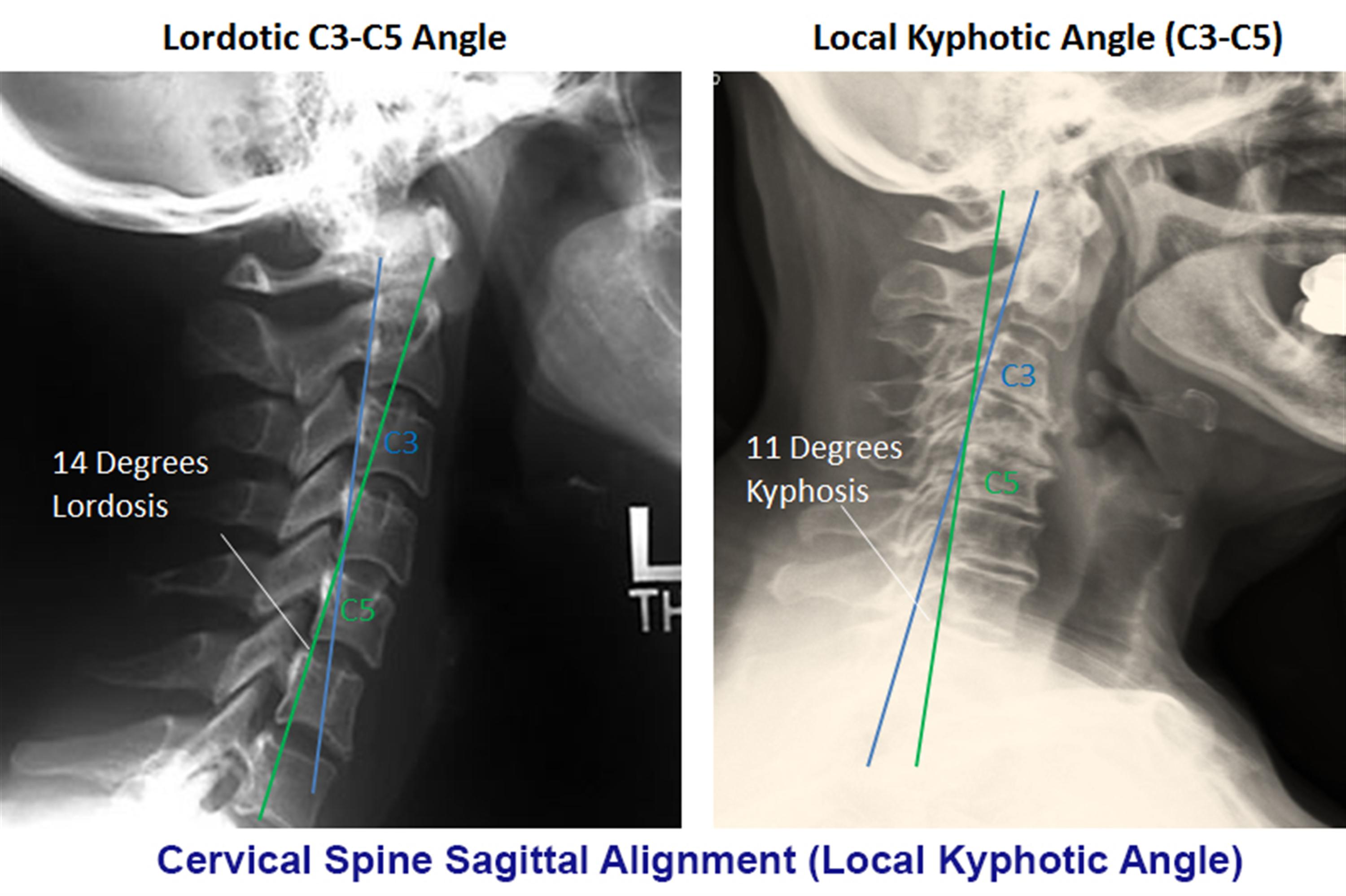
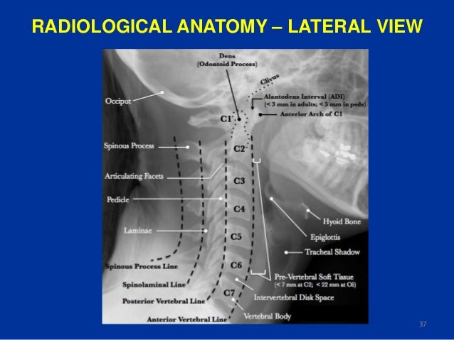
Cervical spine xray anatomy. As viewed from the side the cervical spine forms a lordotic curve by gently curving toward the front of the. Although called the odontoid peg. It normally consists of seven vertebrae.
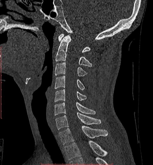
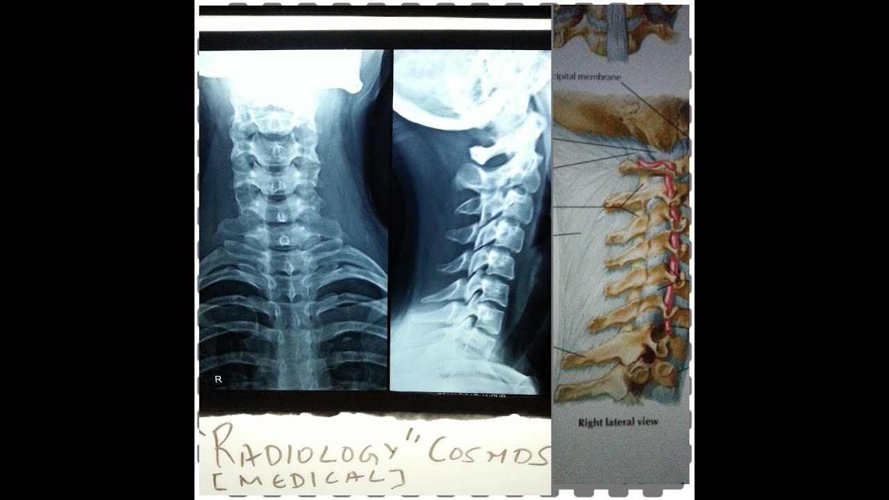
We would like to show you a description here but the site wont allow us. This page was last updated on 1698. The lateral view is often the most informative image.
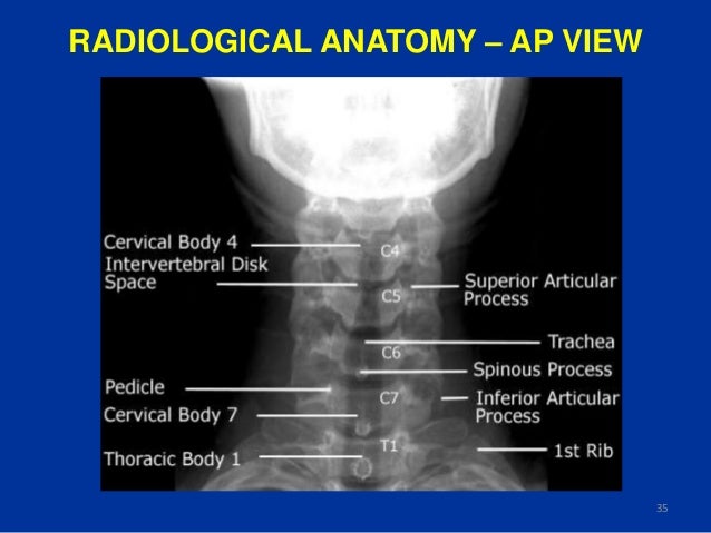
Cervical spine radiographs are often used to confirm that an anterior fixation cervical spine fusion performed with interposition grafts remains stable and secure. The cervical spine is the upper part of the spine extending from the skull base to the thorax at the level of the first vertebra with a rib attached to it. The anteroposterior radiograph anterior aspect shows the vertebral bodies of five lumbar vertebrae their transverse processes spinous and upper and lower joints.
The top of the cervical spine connects to the skull and the bottom connects to the upper back at about shoulder level. Its main function is to support the skull and maintain the relative positions of the brain and spinal cord. Radiological anatomy of the lumbar spine.
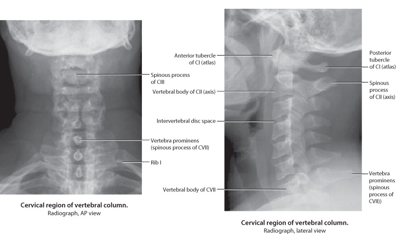
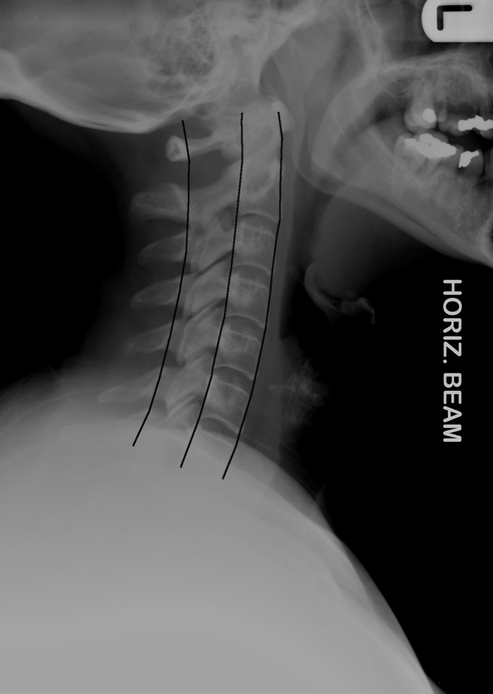
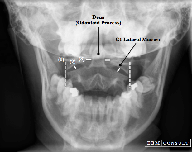
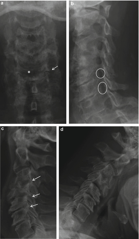
Line 3 will sometimes show a slight step at the c2 level particularly in children 8. 1997 michael l. Views of the cervical spine.
Trace these lines along the full length of the cervical spine. Understanding x rays of the cervical spine this videounderstanding x rays of the cervical spineis designed for the practitioner who has access to x rays for diagnosis of neck pain and neck disorders see the anatomy section for the anatomy of the cervical spine. Left anterior oblique view.
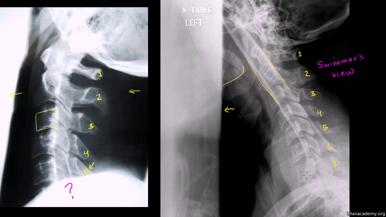
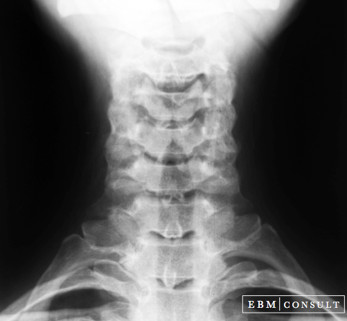
The cervical spine series is a set of radiographs taken to investigate the bony structures of the cervical spine albeit commonly replaced by the ct the cervical spine series is an essential trauma radiograph for all radiographers to understand. Cervical x rays can deliver so much information in so few films. The cervical spine has 7 stacked bones called vertebrae labeled c1 through c7.
Although often less informative than the lateral view this view may nevertheless provide. Odontoid pegopen mouth view. Trauma x ray axial skeleton cervical spine normal anatomy lateral view.
Cervical spine anatomy video.
 B Anteroposterior Axial Cervical Spine X Ray Red Line
 Differential Diagnosis Of Cervical Spine Injuries
 C Spine X Ray Interpretation
 Cervical Spine Ap Lat Anatomy And Physiology Part 10
 Imaging Anatomy Interactive Pacs Like Atlas Of Radiological
 Cervical Spine Radiograph Ap Anatomy Quiz Radiology
 Close Up X Ray Film Show Cervical Spine Or C Spine Neck
 The Cervical Spine
 Normal Cervical Spine Radiographs Radiology Case
 X Ray Spine
 Cervical Spine Radiographic Anatomy Wikiradiography
 Adequacy Of The Lateral Cervical Spine X Ray
 Normal Anatomy
Mri Cervical Spine Sagittal Anatomy
Cervical Spine Radiographic Anatomy Wikiradiography
 X Ray Spine
Xray Of Neck And Cervical Spine Side View Stock Photo Download Image Now
 Interpretations Of The C Spine On Plain Radiography
 Ap Neck Radiograph X Ray
 Normal Anatomy
 Lateral Cervical Spine Radiograph X Ray How To Read
 Body Of Vertebra An Overview Sciencedirect Topics
 The Radiology Assistant Spine Cervical Injury
 Cervical Spine Imaging In Trauma
 Cervical Spine X Ray And 3d Anatomy Case Of Cervical Straightening
Close Up Xray Film Show Cervical Spine Or Cspine Neck Bones
 Cervical Spine Imaging In Trauma
 Cervical Spondylosis Arthritis Of The Neck Orthoinfo Aaos
 Cervical Spine Imaging Normal Anatomy And Degenerative
 Radiology Basics Head Anatomy
 Interpretations Of The C Spine On Plain Radiography


Belum ada Komentar untuk "Cervical Spine Xray Anatomy"
Posting Komentar