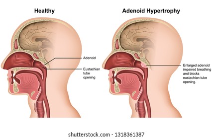
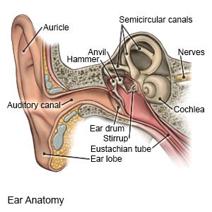
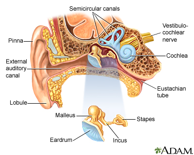
Eustachian Tube Anatomy
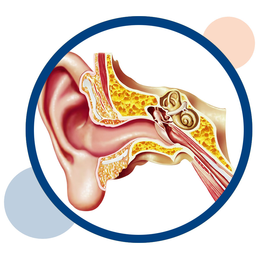
The eustachian tube is a canal that connects the middle ear to the nasopharynx which consists of the upper throat and the back of the nasal cavity. It is a part of the middle ear.
Acclarent Aera Eustachian Tube Balloon Dilation System
The eustachian tube pharyngotympanic tube connects the middle ear cavity with the nasopharynx.
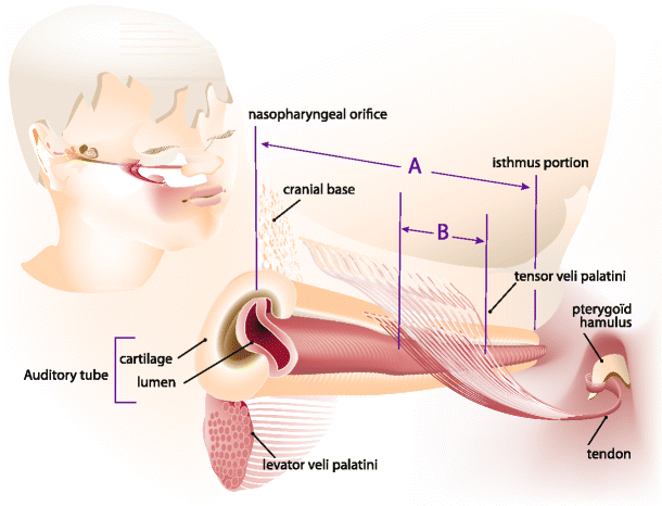
Eustachian tube anatomy. In adult humans the eustachian tube is approximately 35 mm 14 in long and 3 mm 012 in in diameter. The eustachian tube is about 3138 mm 1215 inches long in humans and lined with mucous membrane. The eustachian tube continues from the front wall of the middle ear to the side wall of the nasopharynx progressing along the posterior edge of the medial pterygoid plate.
It aerates the middle ear system and clears mucus from the middle ear into the nasopharynx. The eustachian tube also known as the auditory tube or pharyngotympanic tube is a tube that links the nasopharynx to the middle ear. Eustachian tube also called auditory tube hollow structure that extends from the middle ear to the pharynx throat.
The eustachian tube is located in the para pharyngeal space and is closely linked to the infratemporal fossa. The eustachian tube connects the anterior wall of the middle ear to the lateral wall of the nasopharynx at the level of the inferior nasal concha. It controls the pressure within the middle ear.
It is widest at the pharyngeal opening and tympanic cavity and. In adults the eustachian tube is approximately 43 mm in length and has a variable diameter throughout ie. The tube extends from the petrous portion of the temporal bone to the fleshy lateral wall of the nasopharynx and hence has both bony and cartilaginous elements.
 Hearing Disorders Brain Connection
 The Role Of The Tensor Veli Palatini Muscle In The
 Human Ear Structure And Anatomy Online Biology Notes
 Eustachian Tube Dysfunction Brigham And Women S Faulkner
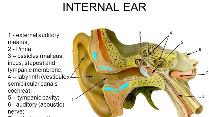
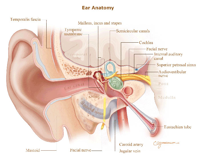
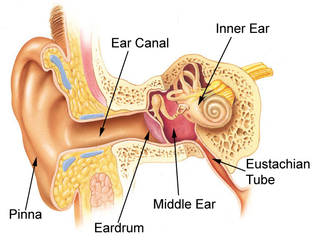
 Anatomy Of The Ear
 Anatomy Of The Human Ear With The Tympanic Membrane The
 Eustachian Tube Dysfunction Pediatric Ent Of Oklahoma
 Eustachian Tube Anatomy Medlineplus Medical Encyclopedia Image
 Eustachian Tube Dysfunction Treatment Surgery Home Remedies
 Eustachian Tube Dysfunction What You Need To Know
![]() Eustachian Pharyngotympanic Tube Anatomy And Function
Eustachian Pharyngotympanic Tube Anatomy And Function

Eustachian Tube Images Stock Photos Vectors Shutterstock
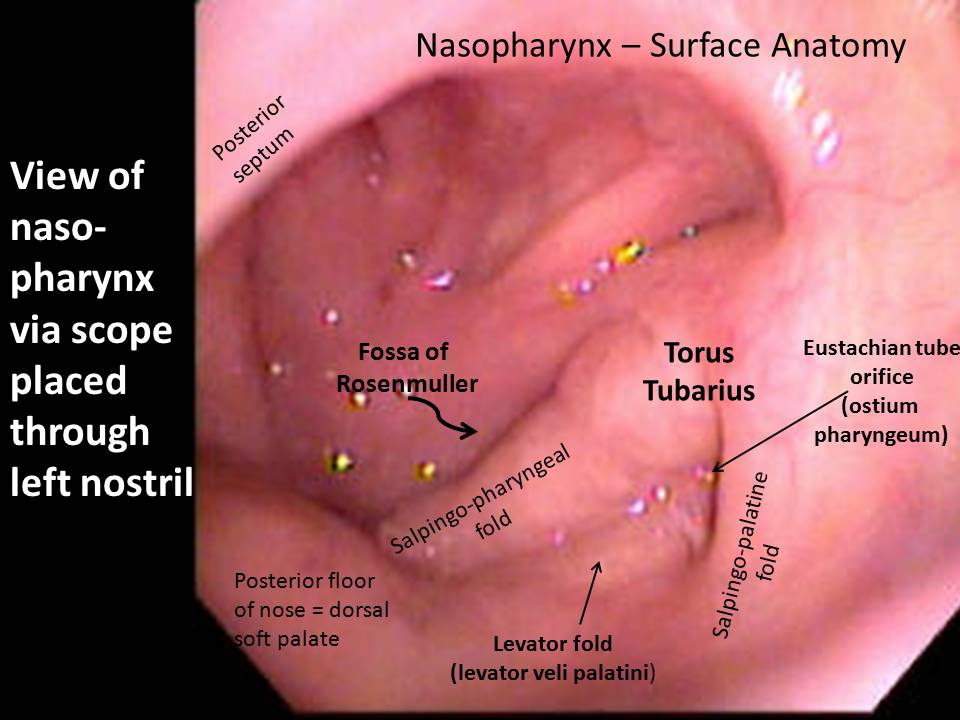
 Eustachian Tube Fossa Of Rosenmuller Anatomy
 Eustachian Tube Images Stock Photos Vectors Shutterstock
 Eustachian Tube Dysfunction Ear Doctor Los Angeles Ca Top
 Eustachian Tube Anatomy 1 Eustachian Tube Dysfunction Ent
 Eustachian Tube Patency Uf Health University Of Florida
 Eystaxianh Salpigga Nornal Anatomy Eustachian Tube From
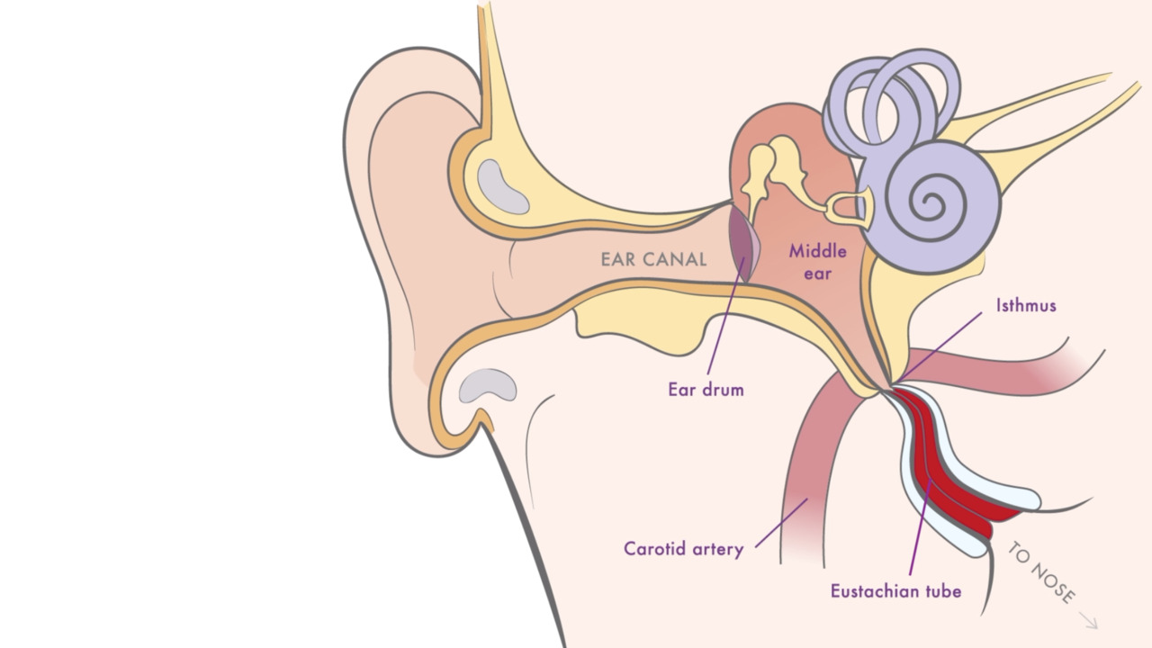
 Eustachian Tubes Auditory Tubes Of The Pharynx
 Patulous Eustachian Tube Management Of The Symptom Of
 How Your Ears Really Work Sinus Relief Center
 Ear Anatomy Outer Ear And Auditory Tube Preview Human Anatomy Kenhub
 Ear Tubes Surgery Placement Procedure In Toddlers Adults
 2 Anatomy Of Ear Msn

 Acclarent Aera Eustachian Tube Balloon Dilation System
How To Tell The Difference Between An Acoustic Neuroma And A
 Eustachian Tube Middle Ear Problems Ashland Audiology
 Figure Nasal Cavity Frontal Sinus Superior
 Why Do My Ears Keep Getting Blocked Quora
![]() Eustachian Pharyngotympanic Tube Anatomy And Function
Eustachian Pharyngotympanic Tube Anatomy And Function
Belum ada Komentar untuk "Eustachian Tube Anatomy"
Posting Komentar