Mca Anatomy
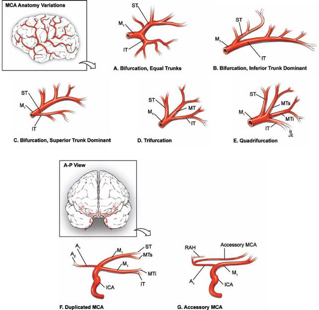
Early branching of the mca bifurcationtrifurcation occurs within 1 cm of its. An accessory mca is a second m1 segment that arises from the a1 anterior cerebral artery aca usually near the anterior communicating artery acoa resembling the recurrent artery of heubner but for the presence of cortical branches fig.
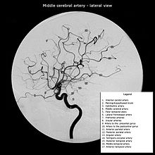
 Middle Cerebral Artery Radiology Reference Article
Return to neuroscience homepage.
Mca anatomy. Mca fenestration is rare with a report incidence of 1. The middle cerebral artery mca is the largest of the three major arteries that channels fresh blood to the brain. Of course the point is that all manner of variations are possible.
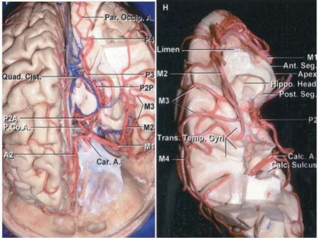
The sphenoidal or horizontal segment. This mca segment perforates parts. Reported incidence of 15 range 02 29.
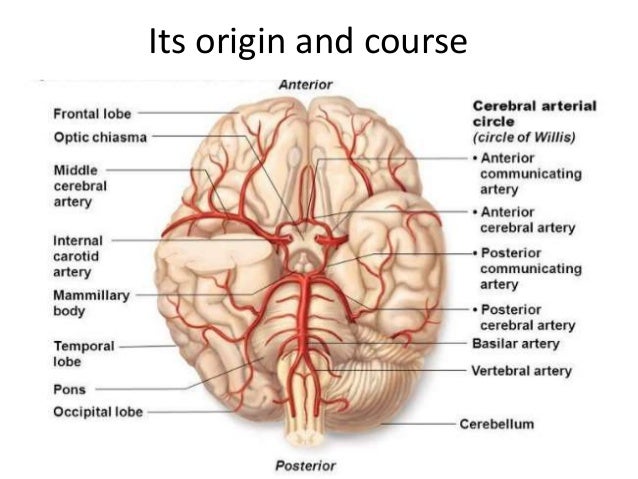
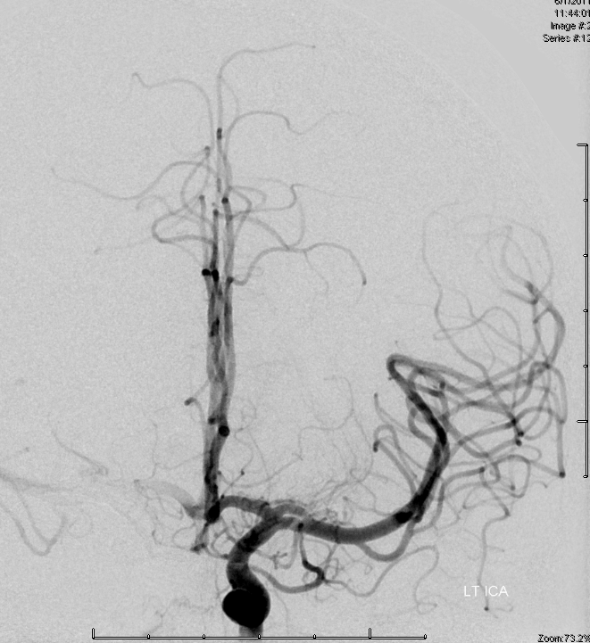
Bridging vein anterior brainstem group cavernous sinus. Move the cursor along the course of the anterior and middle cerebral artery and its branches to identify individual segments and their perfusion targets. At the base of the brain the carotid and vertebrobasilar arteries form a circle of communicating arteries known as the circle of willis.
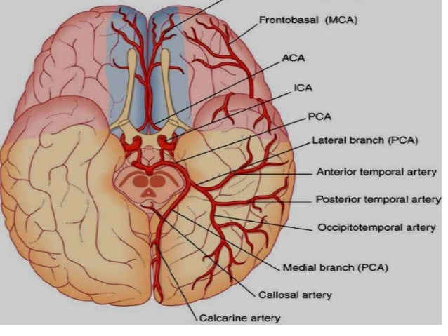
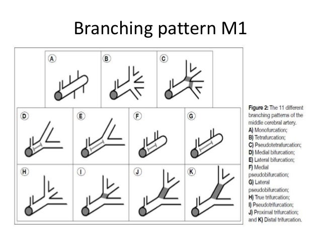
From this circle other arteriesthe anterior cerebral artery aca the middle cerebral artery mca the posterior cerebral artery pcaarise and travel to all parts of the brain. On the opposite end of the silliness spectrum is an mca with no bifurcation. Mca trifurcation this is typically a disposition where each m2 branch takes its lobe frontal parietal or temporal.
Parallels the main mca and supplies the anterior temporal lobe. This segments bifurcates or trifurcates and terminates in the brains cortex. It branches off the internal carotid artery.
4th ventricle and its veins. Variant anatomy mca duplication. Supplies most of the temporal lobe anterolateral frontal lobe and parietal lobe.
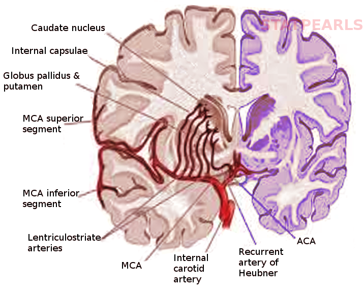
It supplies blood to lateral side. Middle cerebral artery the cortical branches of the mca depicted in red in the diagrams supply the lateral surface of the hemisphere except for the medial part of the frontal and the parietal lobe which is supplied by the aca and the inferior part of the temporal lobe which is supplied by the pca. Stroke m3 aspiration of 1 mm vessel by a 15 mm od catheter.
Middle cerebral artery anatomy the middle cerebral artery mca is one of the three major paired arteries that supply blood to the brain. This segment of the mca also travels through your brain and terminates in. The mca arises from the internal carotid artery ica as the larger of the two main terminal branches mca and anterior cerebral artery and continues into the lateral sulcus where is branches.
Here is a trifurcation no mca bifurcation.
 Mca Anatomy
 Mca Anatomy
 Comparative Cerebrovascular Anatomy Of The Circle Of Willis
 Vasculary Territories Territory Of Middle Cerebral Artery
 Middle Cerebral Artery And Branches Alila Medical Images
 Middle Cerebral Artery Wikipedia
 Internal Carotid Artery An Overview Sciencedirect Topics
 Middle Cerebral Artery Aneurysms Neupsy Key
 Bypass Surgery For Complex Middle Cerebral Artery Aneurysms
 Bypass Surgery For Complex Middle Cerebral Artery Aneurysms
 Middle Cerebral Artery Wikipedia

 Mca Named Segments M1 Etc
 Leptomeningeal Collateral Circulation Wikipedia
Brain Vascular Anatomy With Mra And Mri Correlation
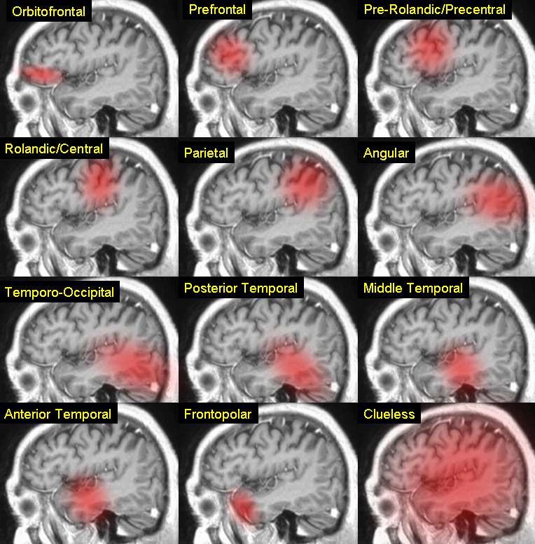
 Aphasia Neurology Medbullets Step 1
 Swiss Medical Weekly Relevance Of The Cerebral Collateral
 Mca Anatomy
 Middle Cerebral Artery Stroke
 Middle Cerebral Artery Radiology Reference Article
Genu Of Middle Cerebral Artery

Belum ada Komentar untuk "Mca Anatomy"
Posting Komentar