Anatomy Chest Cavity
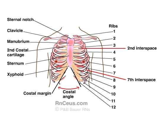
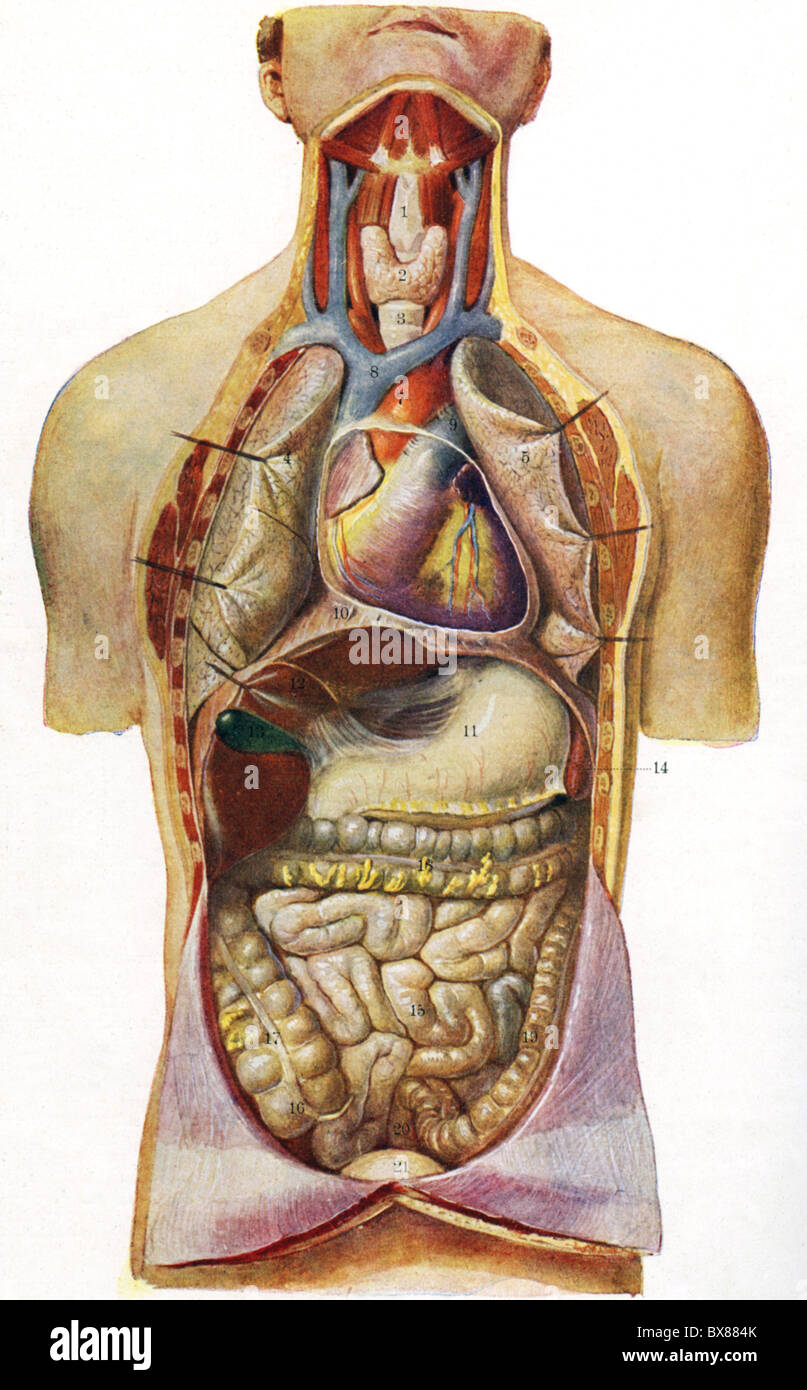
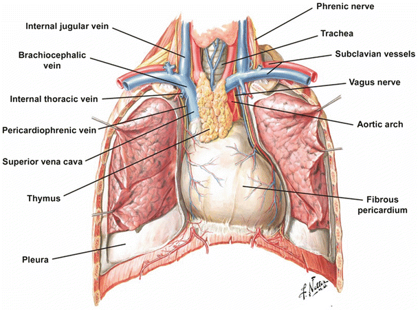
Contains the trachea bronchi lungs esophagus heart and great blood vessels thymus gland lymph nodes and nerve. These sections consist of the manubrium body or corpus and the xiphoid process.
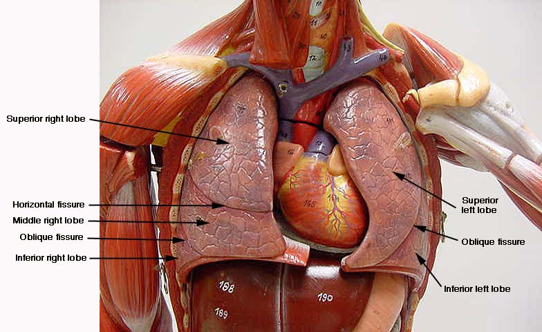
The chest is the area of origin for many of the bodys systems as it houses organs such as the heart esophagus trachea lungs and thoracic diaphragm.
Anatomy chest cavity. The xiphoid process is the small bones seen at the tip of the sternum. Thoracic cavity also called chest cavity the second largest hollow space of the body. The chest cavity is bound by the thoracic vertebrae which connect to the ribs that surround the cavity.
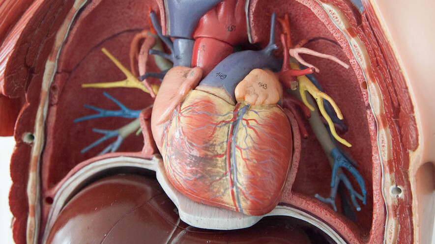
The pleural cavities flank the. The circulatory system does most of its work inside the chest. It contains organs including the heart lungs and thymus gland as well as muscles and various other internal structures.
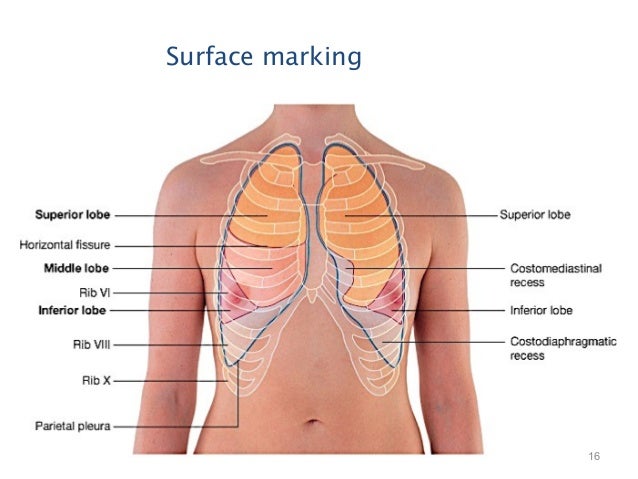
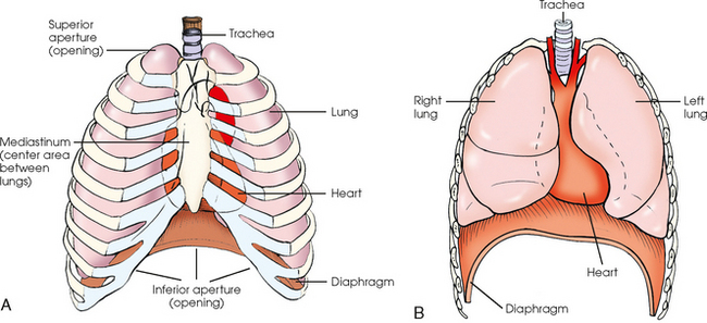
The thoracic cavity is made up of 12 pairs of ribs that connect in the posterior thorax to the vertebral bodies of the spinal column. Diagrams showing the general organisation of the thorax with the pleural cavity and mediastinum anatomy of the lungs histology. In the anterior thorax the first 7 pairs of ribs are attached to the sternum or breastbone by cartilage.
Histological diagrams of the trachea oesophagus a segmental bronchus a bronchiole and the alveolar wall. The thoracic cavity also called the chest cavity is a cavity of vertebrates bounded by the rib cage on the sides and top and the diaphragm on the bottom. Anatomy of the chest cavity and sternum.
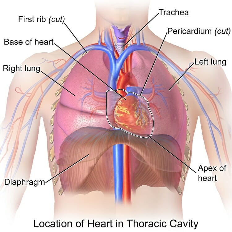
The sternum is made of three sections of bone tissue. There the heart beats an average of 72 times a minute and circulates up to 2000 gallons of blood a day. The thorax includes the thoracic cavity and the thoracic wall.
As well as the following smaller cavities. The thorax or chest is a part of the anatomy of humans and various other animals located between the neck and the abdomen. It is enclosed by the ribs the vertebral column and the sternum or breastbone and is separated from the abdominal cavity the bodys largest hollow space by a muscular and membranous partition the diaphragm.
 Human Anatomy Chest Cavity Anatomy Of Chest Bones Human
 Thoracic Cavity Mediastinum
 Location Of The Heart In The Chest Cavity Thoracic Cavity
Search Anatomy Of The Upper Chest Cavity Right Lateral View
 Thoracic Viscera Radiology Key
 Thoracic Cavity Definition Organs Of Chest Cavity

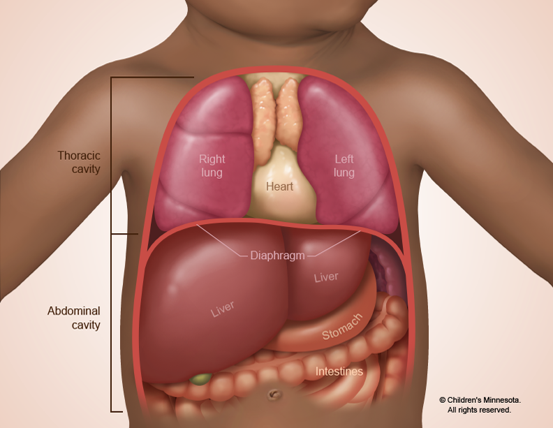 Congenital Diaphragmatic Hernia Treatment Children S Minnesota
 Understanding The Human Chest Thorax Health Life Media

![]() Thorax Anatomy Wall Cavity Organs Neurovasculature
Thorax Anatomy Wall Cavity Organs Neurovasculature
 Chest Cavity
 Thoracic Wall And Breast Illustrations
 Pleural Cavity Wikipedia
 Pulmonary Cavities Anatomy An Essential Textbook 1st Ed
Newsela The Structure And Physiology Of The Heart
 Chest Cavity Shower Curtains Fine Art America

 Anatomy Of The Thoracic Cavity 1st Plastic Model
 Chest Cavity Stock Photos Chest Cavity Stock Images Alamy
 Thoracic Cavity Atlas Of Anatomy
Stock Image Late Victorian Paper Model Showing The Organs
 Thoracic Wall And Breast Illustrations
 Thoracic Cavity Wikipedia
 Anatomy Of The Thoracic Wall Pulmonary Cavities And
 Thoracic Cavity Thoracic Cavity Anatomy Organs
 Anatomy Of The Thoracic Wall Pulmonary Cavities And
 Intro Anatomy 2 Thoracic Cavity

 Amazon Com Professional Medical Anatomy Of Human Organ

Belum ada Komentar untuk "Anatomy Chest Cavity"
Posting Komentar