Bladder And Uterus Anatomy
Bladder vagina uterus fallopian tube ovaries. Anatomy of the female urinary system showing the kidneys ureters bladder and urethra.
 How Well Do Women Know Their Anatomy Viveve Us
Each tube starts with the funnel shaped infundibulum which has fimbriae finger like projections that lie over the ovary.

Bladder and uterus anatomy. It is a potential space prone to fluid collection. Between the uterus and the rectum is the recto uterine space also known as the posterior cul de sac. During pregnancy the uterus takes up significantly more space and severely limits the expansion of the urinary bladder.
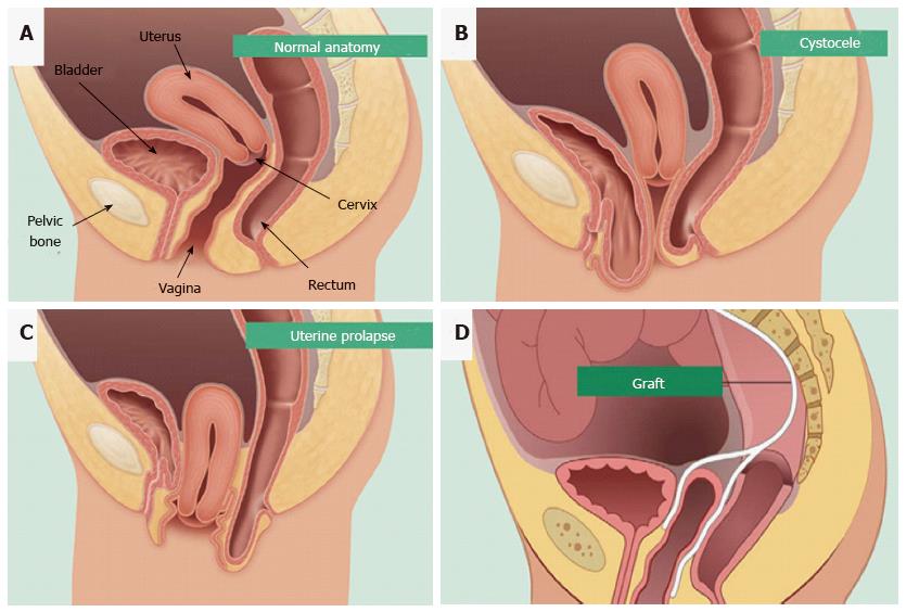
The uterus is also shown. In the normal adult uterus it can be described as anteverted with respect to the vagina and anteflexed with respect to the cervix. This position is referred to as anteversion of the uterus.
The bowel though is more dependent on structural support from the uterus. In most women the long axis of the uterus is bent forward on the long axis of the vagina against the urinary bladder. In females the urinary bladder is somewhat reduced in size and must share the limited space of the pelvic cavity with the uterus that rests superior and posterior to it.
Urine is made in the renal tubules and collects in the renal pelvis of each kidney. Furthermore the long axis of the body of the uterus is bent forward at the level of the internal os with the long axis of the cervix. Anterior to the uterus is the bladder with rectum located posteriorly.
Wear and tear on these supportive structures in the pelvis can allow the bottom of the uterus the floor of the bladder or both to sag through the muscle and ligament layers. Small amounts of physiologic fluid accumulate during ovulation and menses. The urine flows from the kidneys through the ureters to the bladder.
The exact anatomical location of the uterus varies with the degree of distension of the bladder. The female pelvic organs. The uterus and the bladder are held in their normal positions just above the inside end of the vagina by a hammock made up of supportive muscles and ligaments.
Webmds bladder anatomy page provides a detailed image and definition of the bladder and describes its function location in the body and conditions that affect the bladder. The ampulla is the widest part of the tube which narrows to form the isthmus. The urine is stored in the bladder until it leaves the body through the urethra.
Some women develop bladder prolapse whether or not they undergo hysterectomy but rectocele is a consequence of hysterectomy for the majority of hysterectomized women. The bladder is supported by anatomical structures in addition to the structural support it gets from the uterus. The uterine tubes lie between the ovaries and uterus in the broad ligament.
 The Urinary Bladder Human Anatomy
 Anatomy Location Of Bladder Vaginal Canal Cervix And
 Bladder Anatomy Overview Gross Anatomy Microscopic Anatomy
 Stock Illustration Female Bladder With Uterus Anatomy
 Placenta Accreta Overview Brigham And Women S Hospital
 Figure Pelvic Exam A Doctor Or Pdq Cancer
 Bladder Sling Complications Pain Mesh Erosion Perforation
 Uterine Cancer General Information Symptoms Signs Of
 Uterine Sarcoma Treatment Mhealth Org
 Multimodality Imaging Of Pelvic Floor Anatomy Springerlink
 Bladder Cancer Treatment Mhealth Org
 The Uterus Structure Location Vasculature Teachmeanatomy
 Rectocele Diagram Surgery Female Genital Anatomy Images
 Extraperitoneal Retroperitoneal Subperitoneal
 Muscle Invasive Bladder Cancer Symptoms Diagnosis
 Uterine Prolapse During Pregnancy Everything You Need To Know
 How To Treat Pelvic Organ Prolapse Inclusive Care Llc
 Pelvic Organ Prolapse What Is Pelvic Organ Prolapse Best
 About Your Bladder The Facts Continence Foundation Of
 12195 01xv2 Pelvic Structures Anatomy Exhibits
 Pelvis Clinical Anatomy A Case Study Approach
 Uterus Ovaries And Bladder Posterior Canvas Print
 Endometrial Mesenchymal Stem Cells As A Cell Based Therapy
 Solved Where Is The Uterus With Respect To The Urinary


Belum ada Komentar untuk "Bladder And Uterus Anatomy"
Posting Komentar