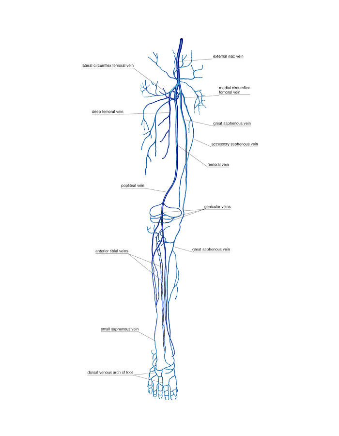
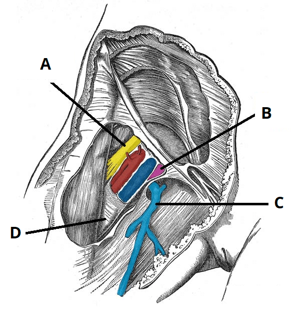
Femoral Vein Anatomy
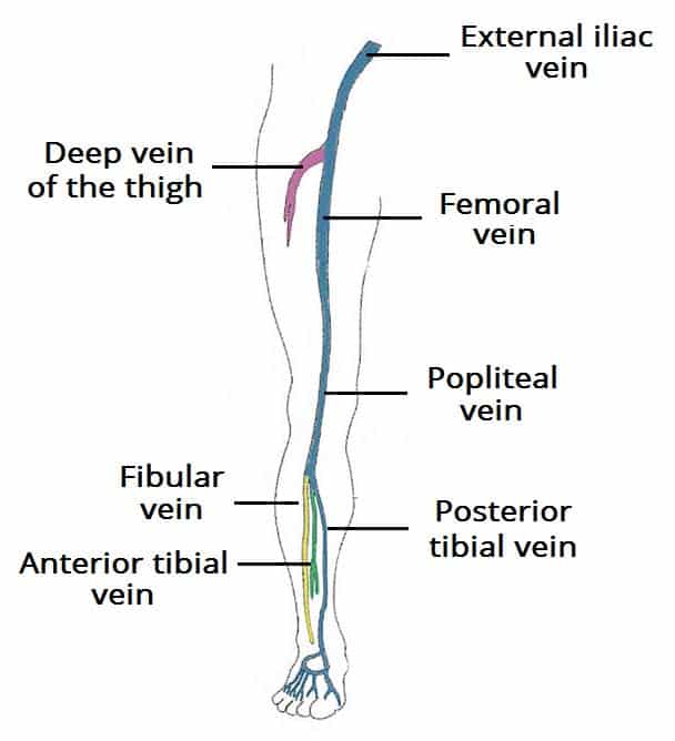
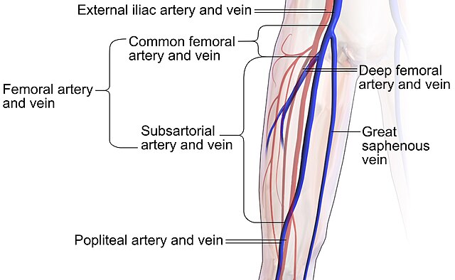
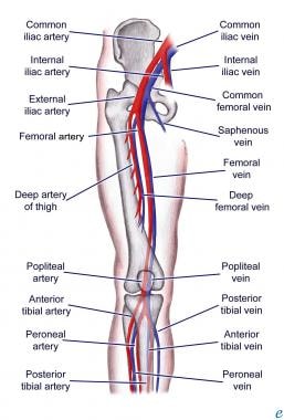
Femoral vein anatomy continuation of the popliteal vein lies in the intermediate compartment of the femoral sheath accompanies the femoral artery in the femoral triangle at the inguinal ligament it becomes the external iliac vein femoral triangle superior. Therefore it starts at the lower end of the adductor canal ascends in adductor canal and enters the femoral triangle where after traversing the intermediate compartment of the femoral sheath it continues as the external iliac vein behind the inguinal ligament medial to the midinguinal point.
 Anatomy Of The Lower Limb Venous System And Assessment Of
It travels in close proximity to the femoral artery.

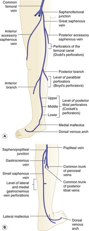
Femoral vein anatomy. Deep vein of the thigh whose entry marks the border between the subsartorial vein and common femoral vein. Several large veins drain into the femoral vein. Great saphenous vein into the common femoral vein.
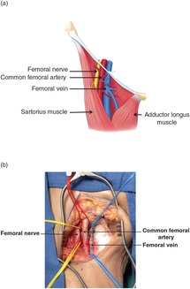
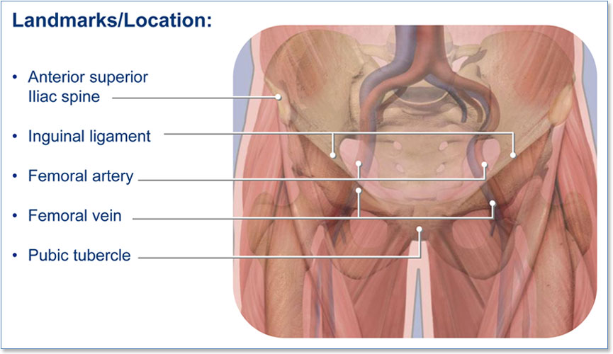
This vein is one of the larger vessels in the venous system. Sartorius crossing the adductor longus muscle roof. Inguinal ligament medial border.
Proximal to the confluence with the deep femoral vein the femoral vein is commonly known as the common femoral vein. Instead of draining deoxygenated blood from specific parts of the body it receives blood from several significant branches. These include popliteal the profunda femoris and the great sapheneous veins.
The femoral vein lies to the midline side of the femoral artery and is considered a continuation of the popliteal vein. Adductor longus lateral border. It begins at the gap of the adductor magnus muscle an inner thigh muscle and the femur.
It becomes the external iliac vein as it ascends posterior to the inguinal ligament. In the distal adductor canal the vein is posterolateral to the superficial femoral artery. Popliteal vein becoming the subsartorial or superficial femoral vein at the adductor.
The femoral vein is the upward continuation of the popliteal vein in the adductor hiatus.
 Lower Extremity Venous Anatomy Dallas Tx Venous System
 Vasculair Intervention Startradiology

 Femoral Artery Injuries Chapter 35 Atlas Of Surgical
 Venous Drainage Of The Lower Limb Teachmeanatomy

 Cross Section Anatomy Transverse Axial Hip Buttocks
 Varicosities Affecting The Lower Limb Veins Consequent To A


Illustration Of Left Spermatic Vein Access From The Right
 Venous System Of The Lower Limb
 Section 2 Anatomy And Physiology
 Vascular Access Tintinalli S Emergency Medicine A
 Varicose Vein Anatomy Pathophysiology Managemant
 Femoral Vein Wikiwand
 Femoral Vein Wikipedia
 External Iliac Vein An Overview Sciencedirect Topics
 Femoral Vein Stock Photos Femoral Vein Stock Images Alamy
 Vein Of Lower Limb
 Femoral Vein Stock Photos Femoral Vein Stock Images Page
 Femoral Vein Canvas Prints Fine Art America
 Superficial Femoral Vein Anatomy The Profunda Femoris
 Pa Anatomy Superfical Anterior Thigh Flashcards Quizlet
 Bedside Ultrasonography In Deep Vein Thrombosis
 Limited Vascular Compression Ultrasound For Deep Venous
 Section 2 Anatomy And Physiology
 The Femoral Triangle Borders Contents Teachmeanatomy
 Femoral Artery Cardiovascular System
 Femoral Veins Stock Photos Femoral Veins Stock Images Alamy
Belum ada Komentar untuk "Femoral Vein Anatomy"
Posting Komentar