Horse Stifle Anatomy
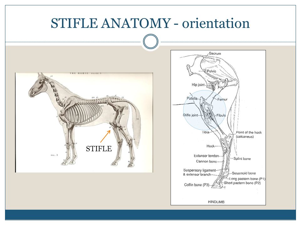
The radiograph at left credit vetwerx is a lateral view of the stifle showing the knee cap or patella and the femur. The stifle is the area where the tibia the bone that forms the gaskin meets the femur the bone that extends upward to the hip.
 Horse Anatomy Mobility Health
When you pick up a horses hind leg the joint bends forward just as your knee does as you climb a staircase.

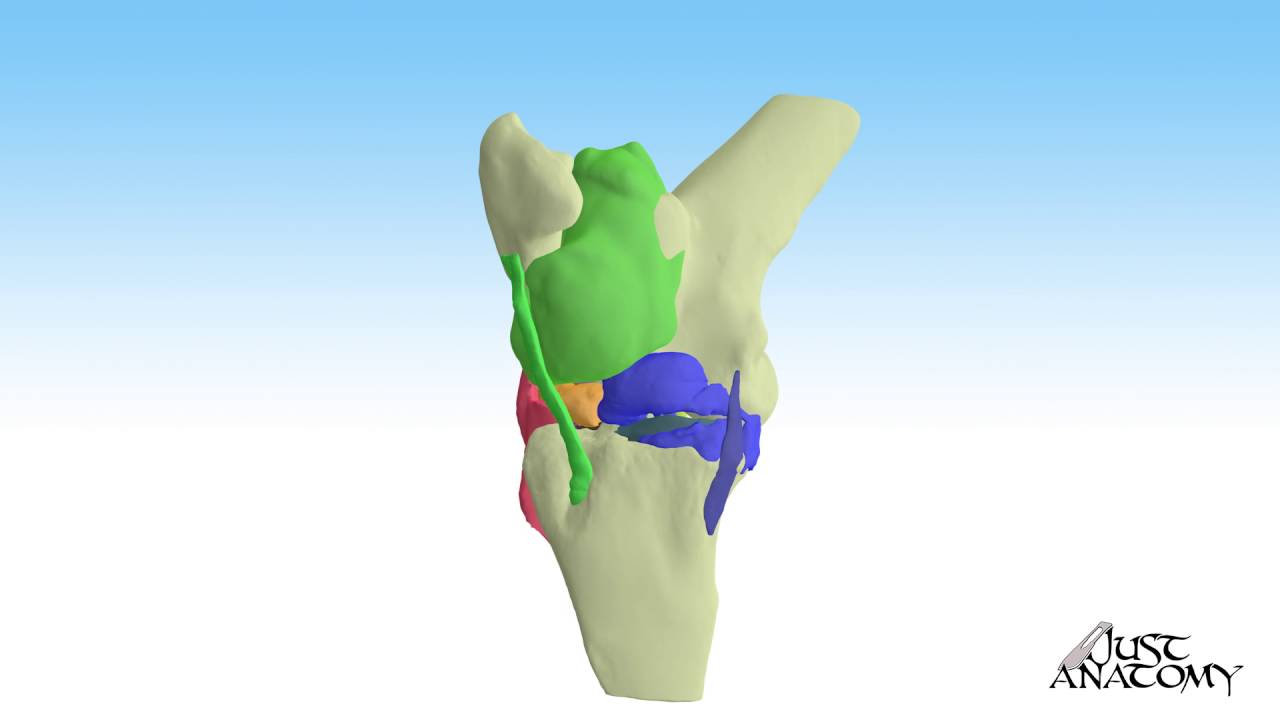
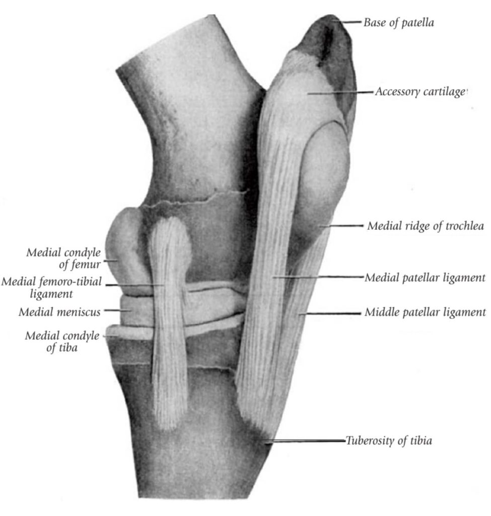
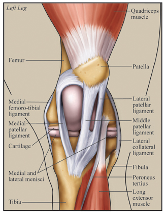
Horse stifle anatomy. Communication of the femoropatellar and medial femorotibial joints has been found 60 to 70 of the time although inflammation anatomic variation and unidirectional flow affect this communication. The stifle is analogous to the human knee. The bones that make up the stifle are the femur thigh tibia shin and patella kneecap.
There are various disease processes that affect the nature of the synovial fluid because of inflammation and disease in the synovial membrane. Femoropatellar medial femorotibial and lateral femorotibial. As the leg moves the patella rides up and down the trochlear ridges of the femur in the femoropatellar joint.
Observe your horse to see if it holds its leg taut and if it drags the toes of its hoof on the ground behind it. The synovial membrane secretes the synovial fluid which provides lubrication within the joint. The equine stifle consists of three compartments.
A horse with a locked stifle will likely hold its hind leg stiff and straight unable to unlock the joint. The mother of all joints. The stifle lifts the leg upward and forward so its pretty critical to moving.
Stifle anatomy radiograph patella femur the equine stifle corresponds to the human knee. The horses stifle is akin to a human knee and it usually bends forward. Anatomy the stifle is the equivalent of the human knee and it is the largest most complex joint in the horse.
Inflammation in the joint causes excessive fluid production.
 Understand Stifle Stresses In Dressage Horses Dressage Today
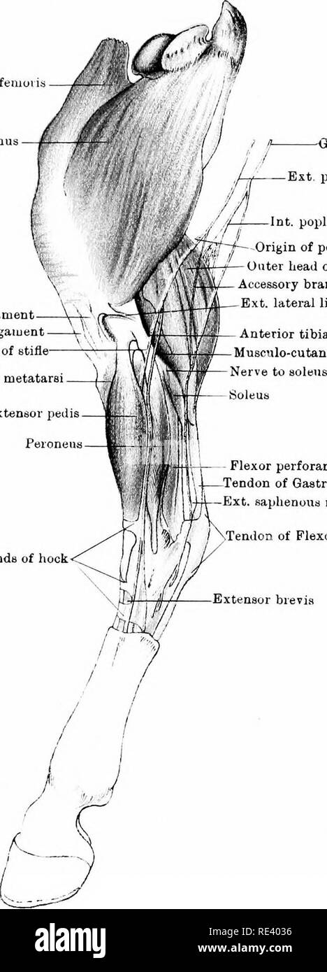 The Anatomy Of The Horse A Dissection Guide Horses Viii
How To Perform Arthrocentesis Of The Compartments Of The
How To Perform Arthrocentesis Of The Compartments Of The
 Equine Stifle Joint
 Equine Stifle In Parker Berthoud Boulder Co Vetwerx Equine
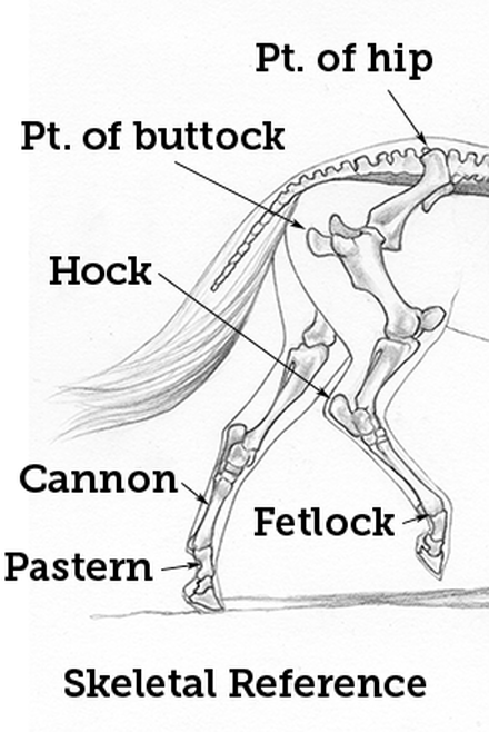
 Conformation Equicessities
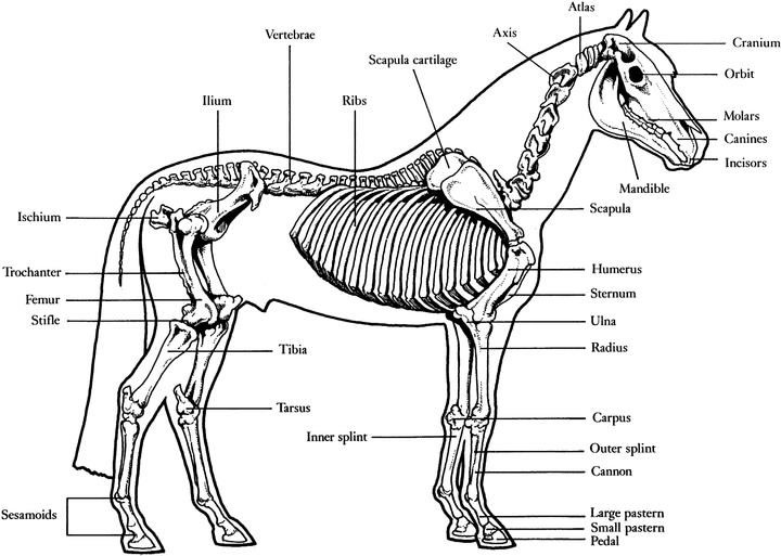 Horse Vertebrate Anatomy Diagram Of A Horse

 Parts Of A Horse Useful Horse Anatomy With Pictures 7 E S L
 Regional Anesthesia In Equine Lameness Musculoskeletal
 Novobrace Tendonitis Desmitis And Soft Tissue Injury
 3 Steps To Stronger Stifles Expert How To For English Riders

:max_bytes(150000):strip_icc()/GettyImages-476732325-56d75b453df78cfb37dadb81.png) How To Treat Locked Stifle Joints In Horses
How To Treat Locked Stifle Joints In Horses
 Horse Hock Massage For Better Performance Expert How To
 Importance Of Proper Hind Leg Conformation Equimed Horse

 Locking Stifles What Does It Mean Darling Downs Vets
 How Do We Diagnose Lameness In Your Horse Ppt Video
 Locking Stifles Henderson Equine Clinic
 The Chronicle Of The Horse
Straight From The Horse S Muscles Tension In The Tensor Muscle
 Hindlimb Anatomy Physiology Wikivet English
 A New Approach To Stifle Injuries The Horse Owner S Resource
:max_bytes(150000):strip_icc()/hind-leg-problems-in-horses-1886457_FINAL-5bf466b446e0fb0051770a3e.png) Hind Leg Problems In Horses Causes And Treatment
Hind Leg Problems In Horses Causes And Treatment
 Equine Stifle An Overview Sciencedirect Topics
 Stifle Joint Anatomy Of The Dog On Mri
 Horse Anatomy Mobility Health
 Horse Anatomy I Mikki Senkarik
 Equine Stifle An Overview Sciencedirect Topics
Belum ada Komentar untuk "Horse Stifle Anatomy"
Posting Komentar