Pelvic Muscle Anatomy Ct
As such you can also divide the musculature that moves the thigh at the hip joint into quadrants. This mri male pelvis axial cross sectional anatomy tool is absolutely free to use.
 Abdominal Ct Anatomy Radiology Key
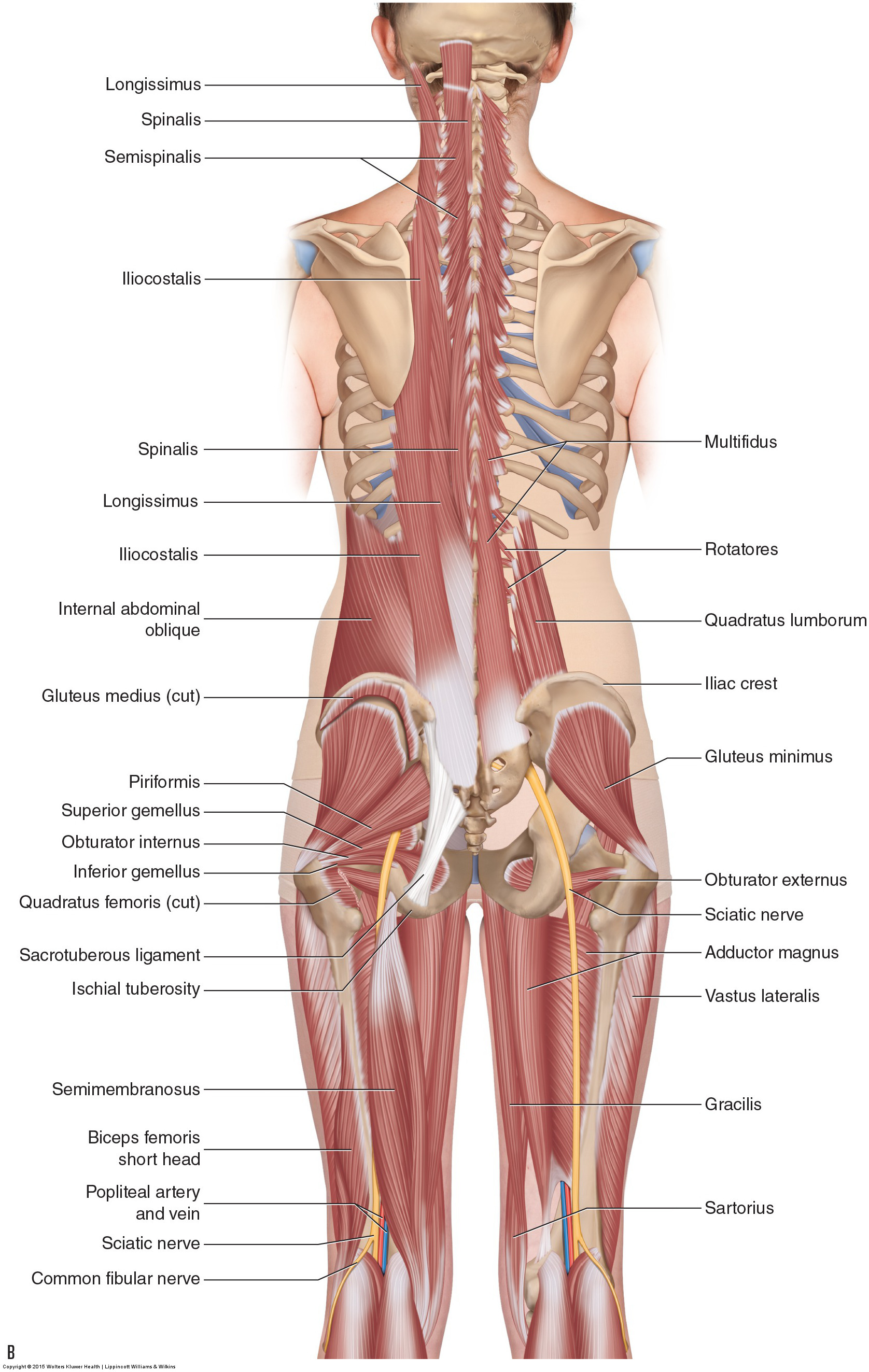
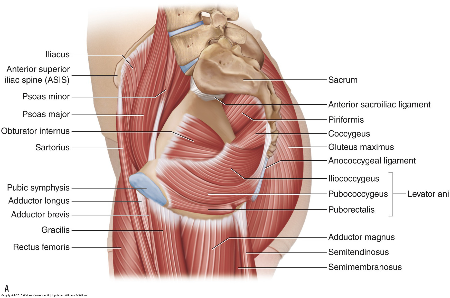
The pelvic floor muscles.

Pelvic muscle anatomy ct. Anatomy ct axial abdomen and pelvis male male abdomen and pelvis ct scan form no 1. Talos i f jakab m kikinis r. Pelvic muscles that cross the hip joint and attach onto the thighleg muscles that cross the hip joint are usually thought of with respect to their open chain motion of the thigh relative to the pelvis at the hip joint.
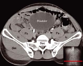
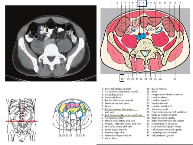
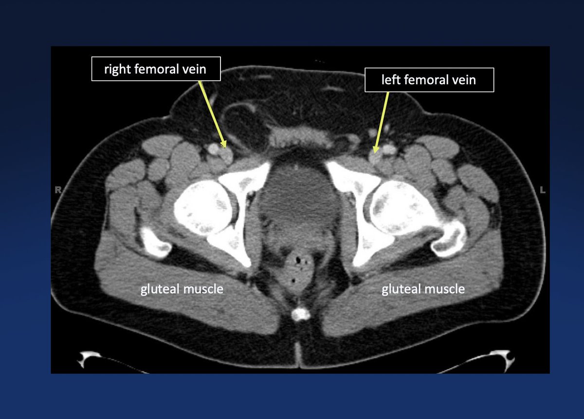
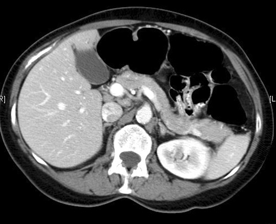
Ct during the administration of intravenous contrast gives excellent cross sectional information about the vessels in the pelvis and their relationship to surrounding structures. 2 psoas muscle 4 sacrum 6 obturator internus muscle 13 ureter 14 bladder 22 small bowel. Ct anatomy of the pelvis.
Learn the diagnosis of ct and methods of computed tomography. By teachmeseries ltd 2019. The arteries have a round even calibre whereas the veins are usually larger and more oval shaped in the supine position.
Use the mouse scroll wheel to move the images up and down alternatively use the tiny arrows on both side of the image to move the images on both side of the image to move the images. Atlas of ct anatomy of the abdomen. The pelvic floor is also known as the pelvic diaphragm.
In this article we shall look at the anatomy of the muscles that make up the inferior lining of the cavity. Anatomy of the abdomen and male pelvis using cross sectional imaging ct interactive atlas of human anatomy we have created an anatomical atlas of abdominal and pelvic ct which is an interactive tool for studying the conventional anatomy of the normal structures based on a multidetector computed tomography. Pelvic muscles ct anatomy and ct scan of the abdomen and pelvis shows a normal appendix 7 pelvic muscles ct anatomy pelvic muscles ct anatomy and ct scan of the abdomen and pelvis shows a normal appendix gallery at human diagram chart.
15 liver 16 oesophagus 17 stomach. We shall look at the individual roles of these muscles their innervation and blood supply and any clinical correlations. This photo gallery presents the anatomy of the abdomen by means of ct axial coronal and sagittal reconstructions.
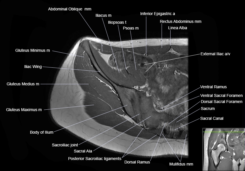
 Mri Pelvis Anatomy Free Male Pelvis Axial Anatomy
 Abdomen And Pelvis Ct
 Sectional Anatomy Of Abdomen
 Pelvis Perineum Anatomy Ppt Download
 Abdominopelvic Cavity And Peritoneum On A Ct
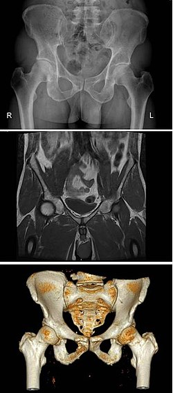
 Mri Hip Anatomy
 Atlas Of Ct Anatomy Of The Abdomen
 Pelvis Wikipedia
 Above Shows A Number Of Possible Measurements Using Mri
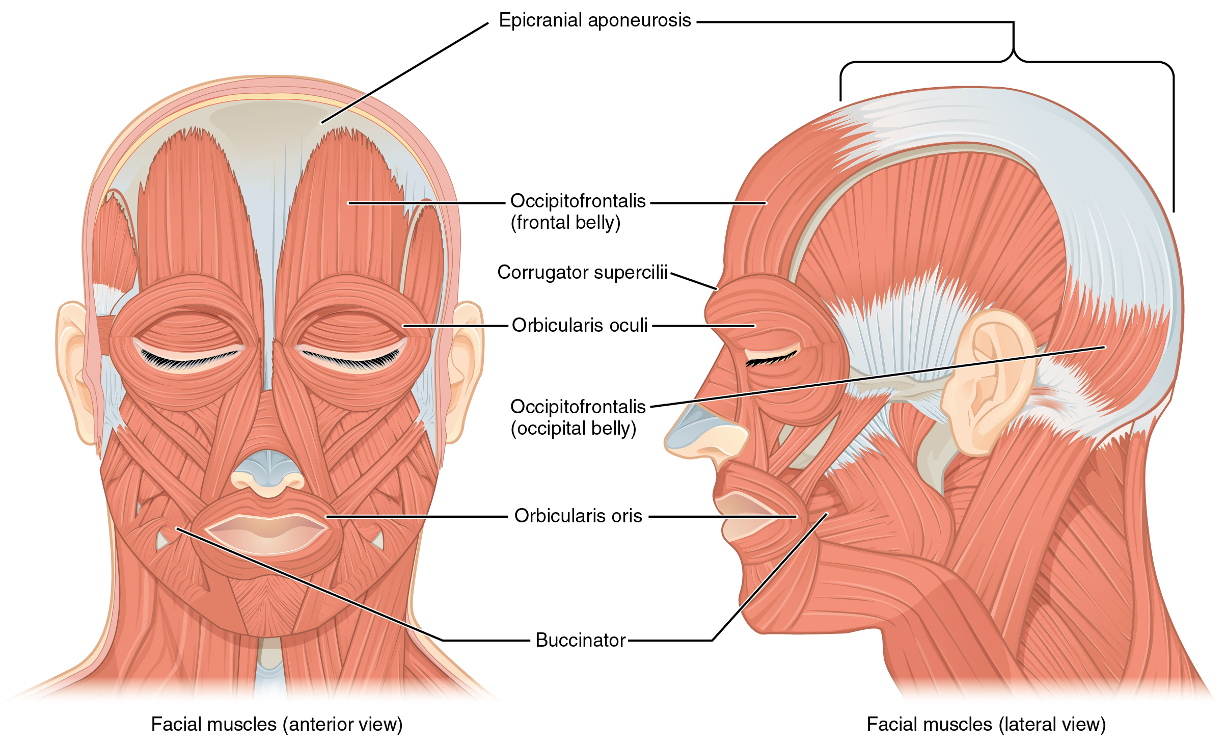 11 3 Axial Muscles Of The Head Neck And Back Anatomy And
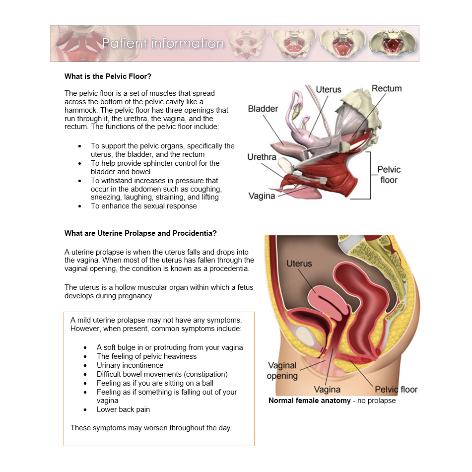 Pelvic Floor Disorders Primal Pictures
 Figure 3 From Ct Anatomy Of The Female Pelvis A Second Look
 X Rays Ct Scans And Mris Orthoinfo Aaos
 The Ct Anatomy Tutor
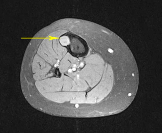
 Obturator Internus Muscle Radiology Reference Article
 Mri Pelvis Anatomy Free Male Pelvis Axial Anatomy
 Elliot K Fishman Ctisus Com On Twitter Ct Pelvic Anatomy
 Axial Ct Image At The Level Of The Piriformis Muscle Open
 The Pelvis Ct Anatomy Mp4
 Anatomy Of The Pudendal Nerve Health Organization For
 Ct Abdomen And Pelvis Coronal Anatomy In The Male
 Muscles Of The Pelvis
 The Ct Anatomy Tutor
 Ecr 2014 C 0356 The Pelvis Revisited A Pictorial Review
 Muscles Of The Pelvis
 Imaging Atlas Of Human Anatomy 4e Pages 151 200 Text
 Gi And Abdomen Radiologic Anatomy
 Startradiology
 Radiology Images
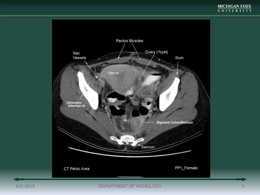
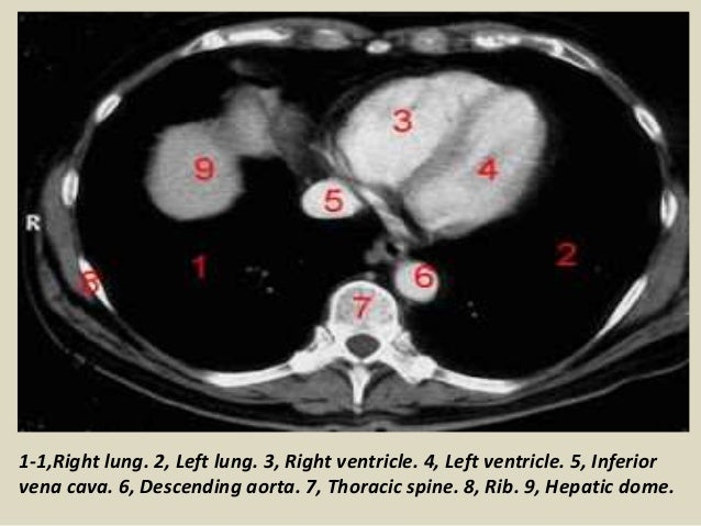
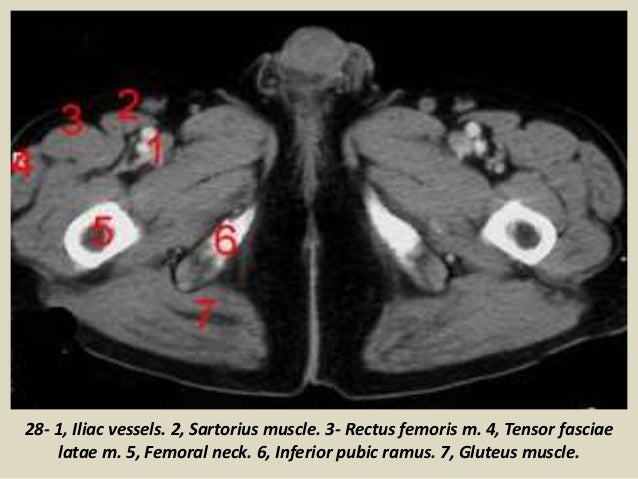 Presentation1 Pptx Ct Normal Anatomy Of The Abdomen And Pelvis
 Presentation1 Pptx Ct Normal Anatomy Of The Abdomen And Pelvis
 Ct Abdomen Anatomy
Body Ct Modules Ct Of The Ovaries And Uterus Ppt Download


Belum ada Komentar untuk "Pelvic Muscle Anatomy Ct"
Posting Komentar