Pelvic Nerves Anatomy
The pelvic plexus lies within the fascia that covers this part of the pelvic wall and floor. Ventral primary rami of spinal nerves s2 s4 sacral plexus inferior rectal n perineal n dorsal n.
 Maternal Anatomy Williams Obstetrics 25e Accessmedicine
Anatomists call the retroperitoneal fascia subserous fascia whereas surgeons refer to this fascial layer as endopelvic fascia.

Pelvic nerves anatomy. There are many organs that sit in the pelvis including much of the urinary system and lots of the male or female reproductive systems. The skin tissues and organs in the pelvis are supplied by the vasculature of the pelvis and innervated by many nerves of the pelvis including the pudendal nerve. This nerve sits in front of the sacral promontory.
Pelvic autonomic nerves include the following. The pelvic venous system is responsible for taking blood from the pelvic walls and viscera back to the main circulation. The pudendal nerve is the main nerve that serves the perineum which is the area between the anus and the genitalia the scrotum in men and the vulva in women.
Like the arterial analogues the external iliac vein primarily drains the lower limbs while the internal iliac vein drains the pelvic viscera walls gluteal region and perineum. Rectal and uterovaginal in the female are in communication with each other. They travel to their sides corresponding inferior hypogastric plexus located bilaterally on the walls of the rectum.
It carries sensory information sensation from the external genitalia and the skin around the anus and perineum. These nerves are a continuation of the lumbar sympathetic trunks. The parasympathetic nerves in the pelvis are the pelvic splanchnic nerves.
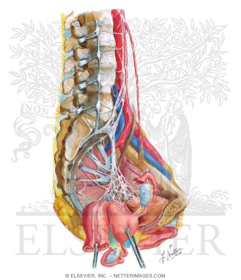
Levator ani muscles including the iliococcygeus pubococcygeus and puborectalis. A small part of the pelvic plexus has been partially dissected out here. The autonomic nerve plexuses of the pelvis prostatic rectal and vesical in the male.
The pelvic ligaments are not classic ligaments but are thickenings of retroperitoneal fascia and consist primarily of blood and lymphatic vessels nerves and fatty connective tissue. From there they contribute to the innervation of the pelvic and genital organs. The pelvic splanchnic nerves arise from the anterior rami of the sacral spinal nerves s2 s4 and enter the sacral plexus.
For updates on video releases follow at accessanatomy on twitter. The perineal nerve innervates muscles of the perineum and pelvic floor. All these nerves sympathetic and parasympathetic are connected to a diffuse and extensive plexus of autonomic nerves called the pelvic plexus.
Veins lymphatics and nerves of the pelvis. These plexuses are formed when the right.
 Pelvic Nerve Anatomy And Lower Urinary Tract Neural Control
 Figure 1 From Diagnosis And Treatment Of Pudendal Nerve
:background_color(FFFFFF):format(jpeg)/images/library/11895/male-pelvic-viscera-and-perineum_english.jpg) Pelvis And Perineum Anatomy Vessels Nerves Kenhub
Pelvis And Perineum Anatomy Vessels Nerves Kenhub
 Anatomy Pelvis Perineum 1 Flashcards Quizlet
:watermark(/images/watermark_5000_10percent.png,0,0,0):watermark(/images/logo_url.png,-10,-10,0):format(jpeg)/images/atlas_overview_image/723/rhPG1aJnnONltuWiHn0awA_nerves-vessels-pelvis-thigh_english.jpg) Pelvic Veins Lymphatics And Nerves Anatomy And Drainage
Pelvic Veins Lymphatics And Nerves Anatomy And Drainage
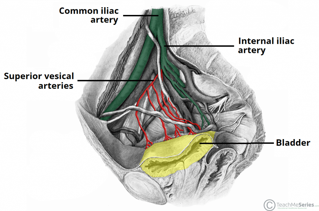 The Urinary Bladder Structure Function Nerves
 Anatomy Of The Pudendal Nerve Health Organization For
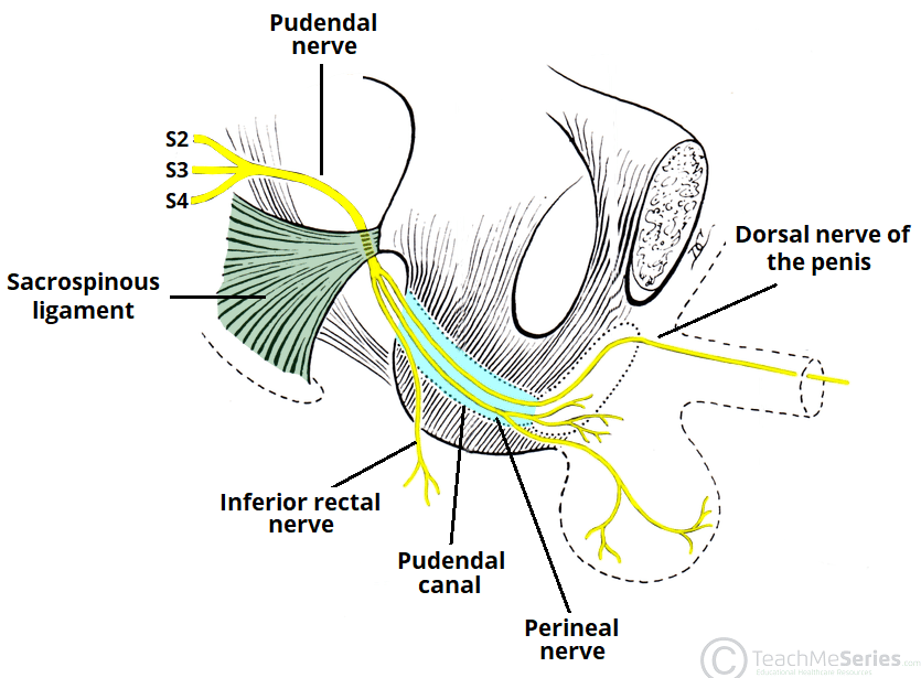 The Pudendal Nerve Anatomical Course Functions
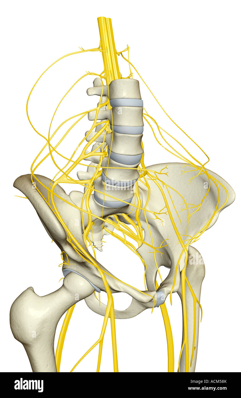 Nerve Supply Of The Pelvis Stock Photo 13185522 Alamy
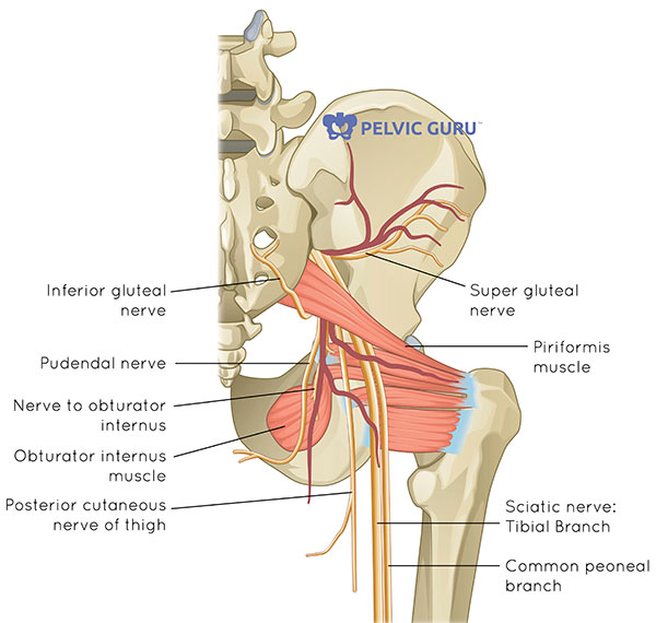 What Is The Pelvic Floor Your Pace Yoga
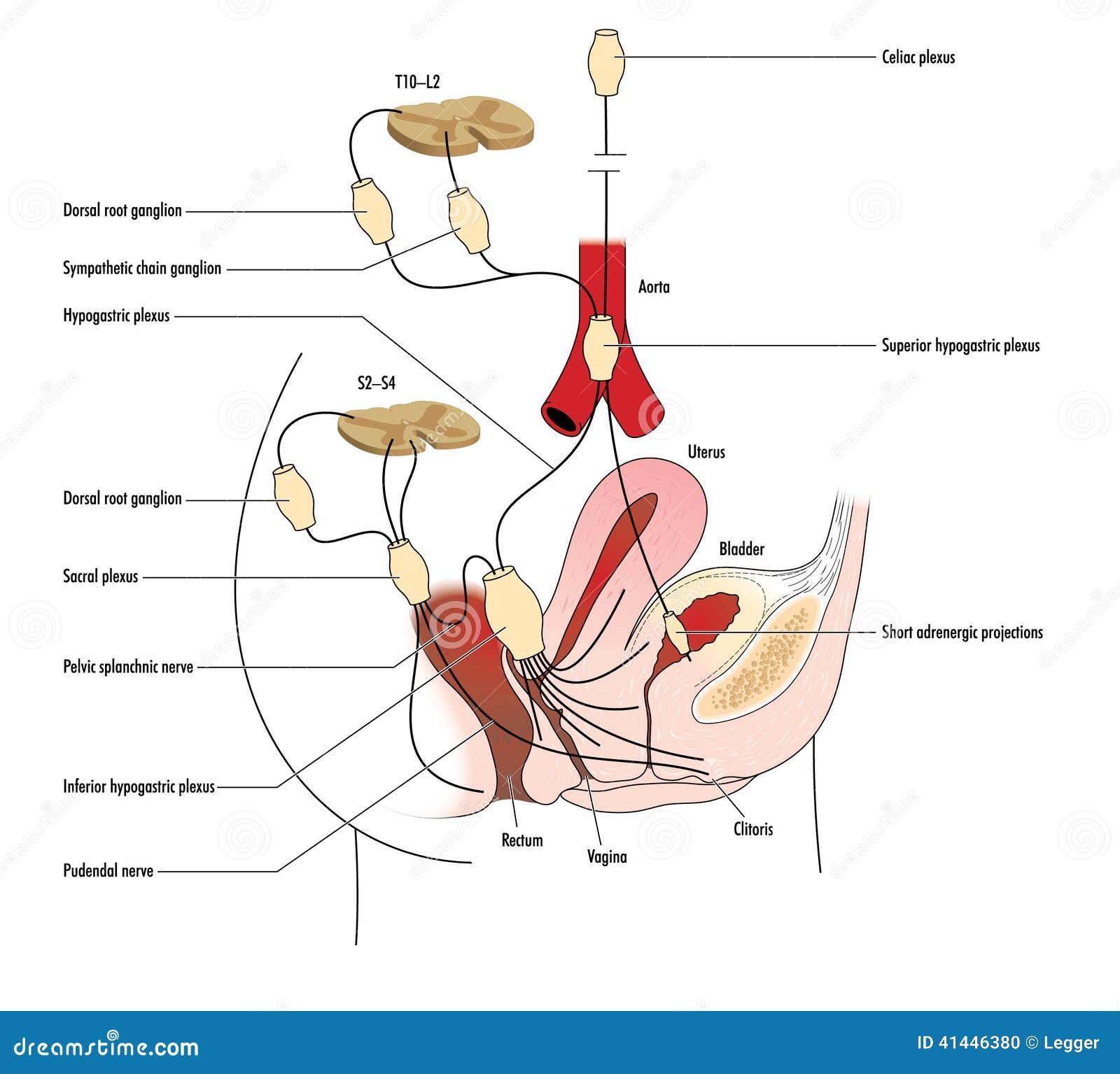 Pelvic Nerves Stock Vector Illustration Of Anatomy Bladder
 Block 2 Lecture 8 Pelvic Neurovasculature Anatomy 1 With
 Module Autonomics Of The Pelvis
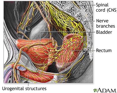 Nerve Supply To The Pelvis Medlineplus Medical Encyclopedia
 11170 03b Nerves Of Male Pelvic Region Anatomy Exhibits
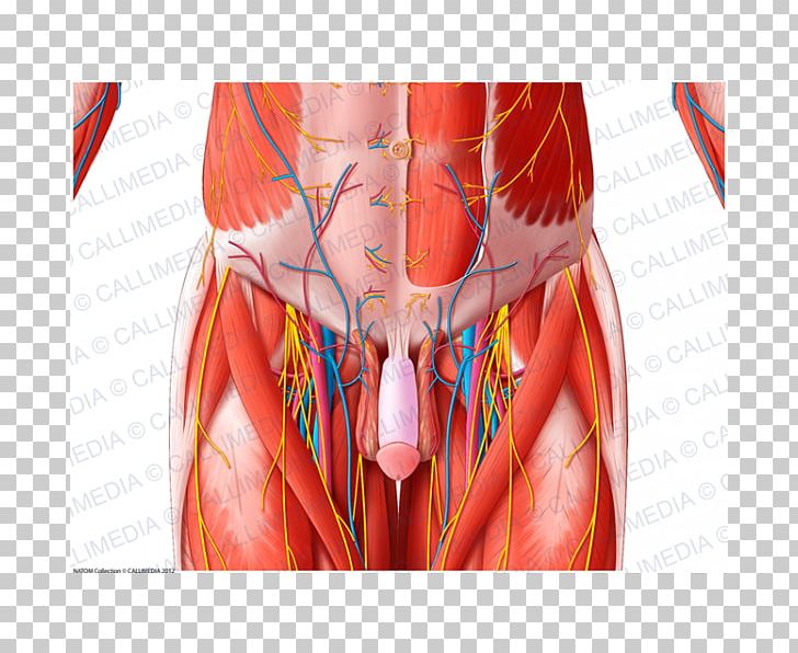 Muscle Blood Vessel Pelvis Nerve Anatomy Png Clipart
 Pelvic Floor Anatomy And Nerves Trivia Questions Quiz
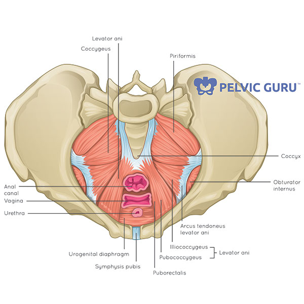 What Is The Pelvic Floor Your Pace Yoga
 Anatomy Perineum Pelvis Brs Part 1 Proprofs Quiz
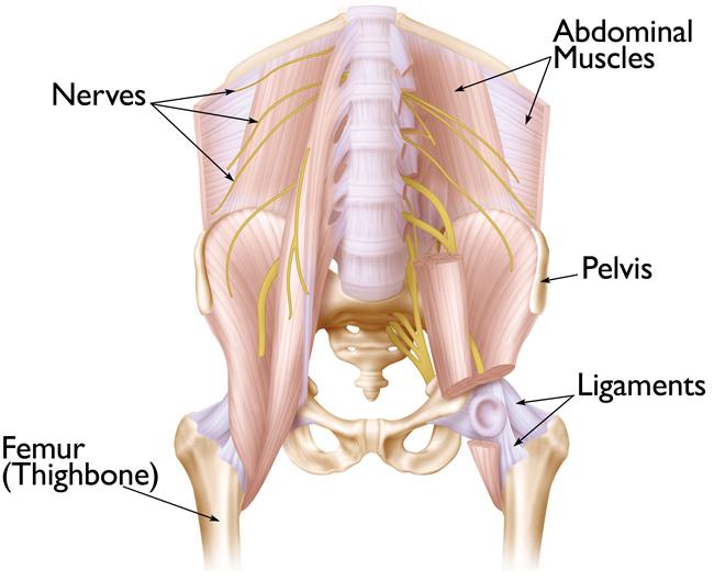 Acetabular Fractures Orthoinfo Aaos
 Surgical Anatomy Of Pelvic Nerves
 Anatomical Feasibility Of Performing Intercostal And
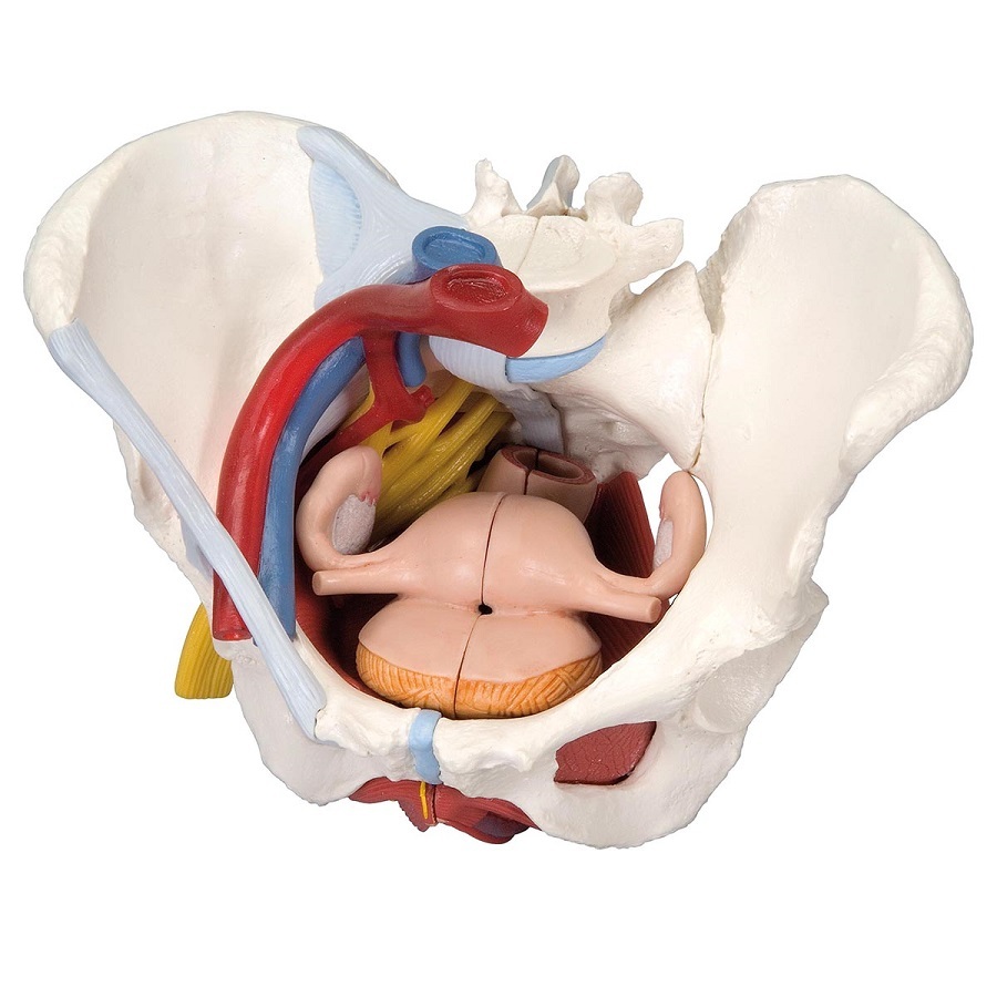 Anatomical Models Of Female Pelvis With Ligaments Vessels


Belum ada Komentar untuk "Pelvic Nerves Anatomy"
Posting Komentar