Posterior Ear Anatomy
Venous drainage is via veins following the arteries listed above. Dorsalis ta dorsal 2.
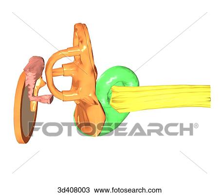 Posterior View Of Inner Ear Anatomy Drawing 3d408003
Often used to indicate the position of one structure relative to another that is nearer the back of the body.
Posterior ear anatomy. It is the vestige of the folded over point of the ear of a remote human ancestor. You will need ear drops but it needs to be prescription. Maxillary artery deep auricular branch supplies the deep aspect of the external acoustic meatus and tympanic membrane only.
Araneae normally have eight eyes in four pairs. Human anatomy denoting the back surface of the body. Helpful trusted answers from doctors.
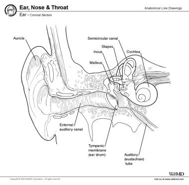
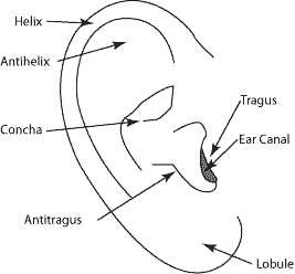
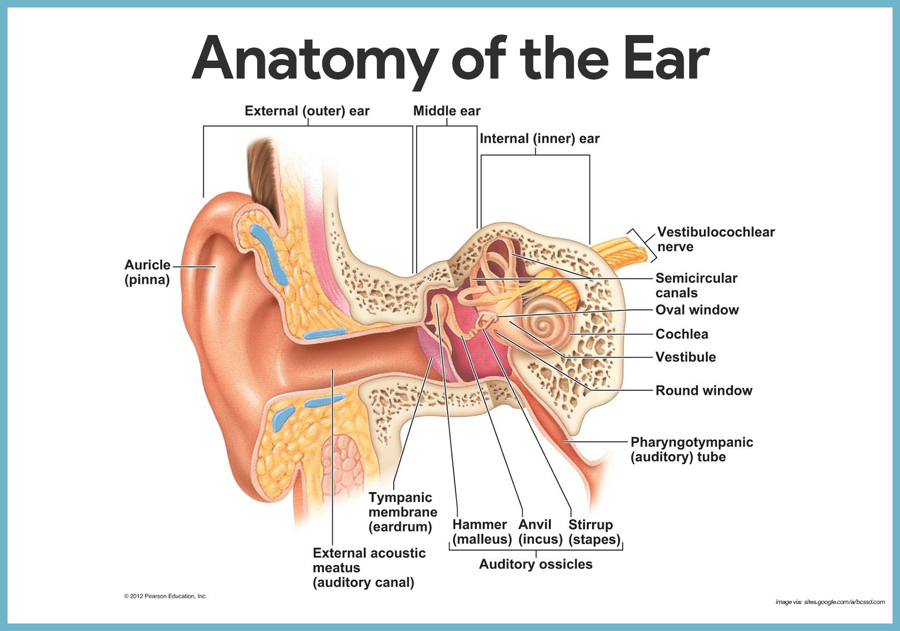
The lobule the fleshy lower part of the auricle is the only area of the outer ear that contains no cartilage. Sound funnels through the pinna into the external auditory canal a short tube that ends at the eardrum tympanic. Variant anatomy of the external ear can be divided into congenital and acquired entities.
Auricle cartilage covered by skin placed on opposite sides of the head auditory canal also called the ear canal eardrum outer layer also called the tympanic membrane the outer part of the ear collects sound. Do not use q tips or alcohol. Colantino on posterior ear anatomy.
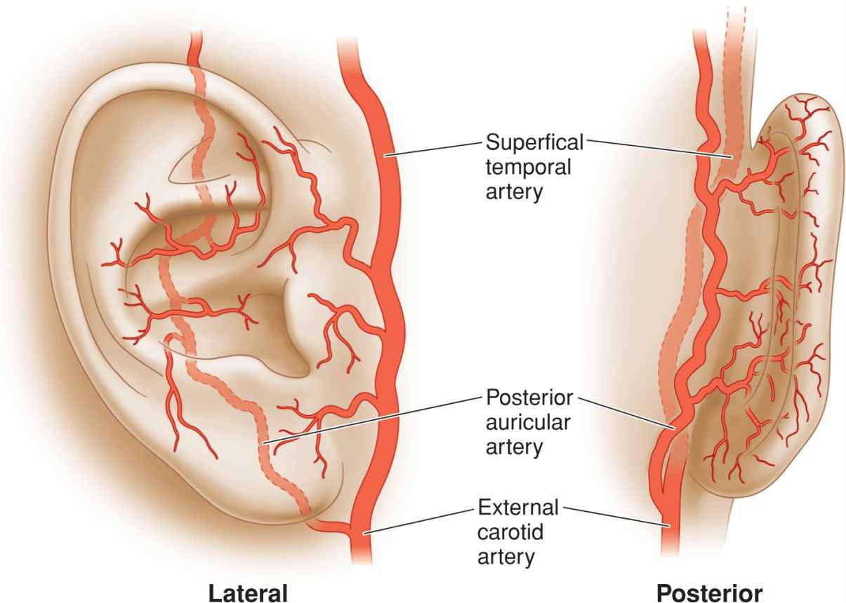
The external ear is supplied by branches of the external carotid artery. The outer ear includes. Acquired entities can further be delineated into intrinsic processes such as cancer and extrinsic processes such as trauma.
The outer ear is called the pinna and is made of ridged cartilage covered by skin. Because of the unusual nature and positions of the eyes of the araneae spiders and their importance in taxonomy evolution and anatomy special terminology with associated abbreviations has become established in arachnology. Posterior pos tēre or directed toward or situated at the back.
Congenital abnormalities of the ear are common and largely affect the shape of the auricle. Sound travels through the auricle and the auditory canal a short tube that ends at the eardrum. In some ears a little prominence known as darwins tubercle is seen along the upper posterior portion of the helix.
Retrolateral refers to the surface of a leg that is closest to the posterior end of an arachnids body.
 Ear Plastic Surgery Key
 Image Result For Anatomy Of Jaw And Ear Muscle Anatomy
/ear-diagram-57bf237f3df78cc16e1dfa27.jpg) Ear Anatomy
Ear Anatomy
 Referred Ear Pain
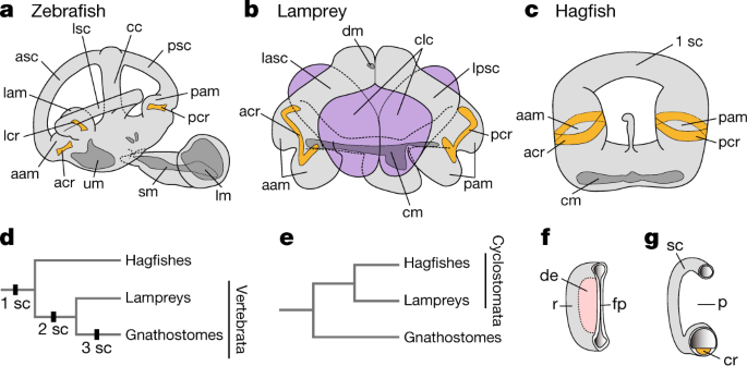 Inner Ear Development In Cyclostomes And Evolution Of The
 Ear Anatomy Overview Embryology Gross Anatomy
 The Posterior Auricular Anatomy Download Scientific Diagram
 Ear Anatomy Outer Ear Mcgovern Medical School
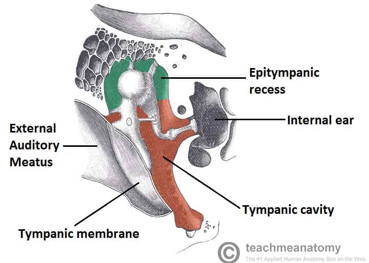 The Middle Ear Parts Bones Muscles Teachmeanatomy
 Basic Human Ear Anatomy And Physiology Outer Middle And
 Ear Disorders Tintinalli S Emergency Medicine A
 Antitragus An Overview Sciencedirect Topics

 Surgical Management Of Skin Cancer And Trauma Involving The
 Middle Ear Anatomy Diagram Quizlet
 Special Senses Anatomy And Physiology Nurseslabs
![]() Ear Anatomy Parts And Functions Kenhub
Ear Anatomy Parts And Functions Kenhub
 Lymphatic Drainage Of The External Ear Semantic Scholar
 The Ear El Oido Anatomy Clipboard Amazon Com Industrial
Anatomy Of Middle Ear With Clinical Correlation Epomedicine
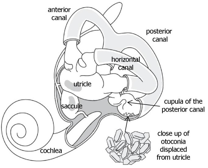 Benign Paroxysmal Positional Vertigo Bppv Vestibular
 Where Are The Major Arteries In Your Ears If You Puncture A

 External Ear
 Figure 1 From A Computational Study Of Ligaments Effect In
 Benign Paroxysmal Positional Vertigo Continuing Professional
Belum ada Komentar untuk "Posterior Ear Anatomy"
Posting Komentar