Posterior Shoulder Anatomy
Pain with jerk test. In human anatomy the shoulder joint comprises the part of the body where the humerus attaches to the scapula the head sitting in the glenoid cavity.
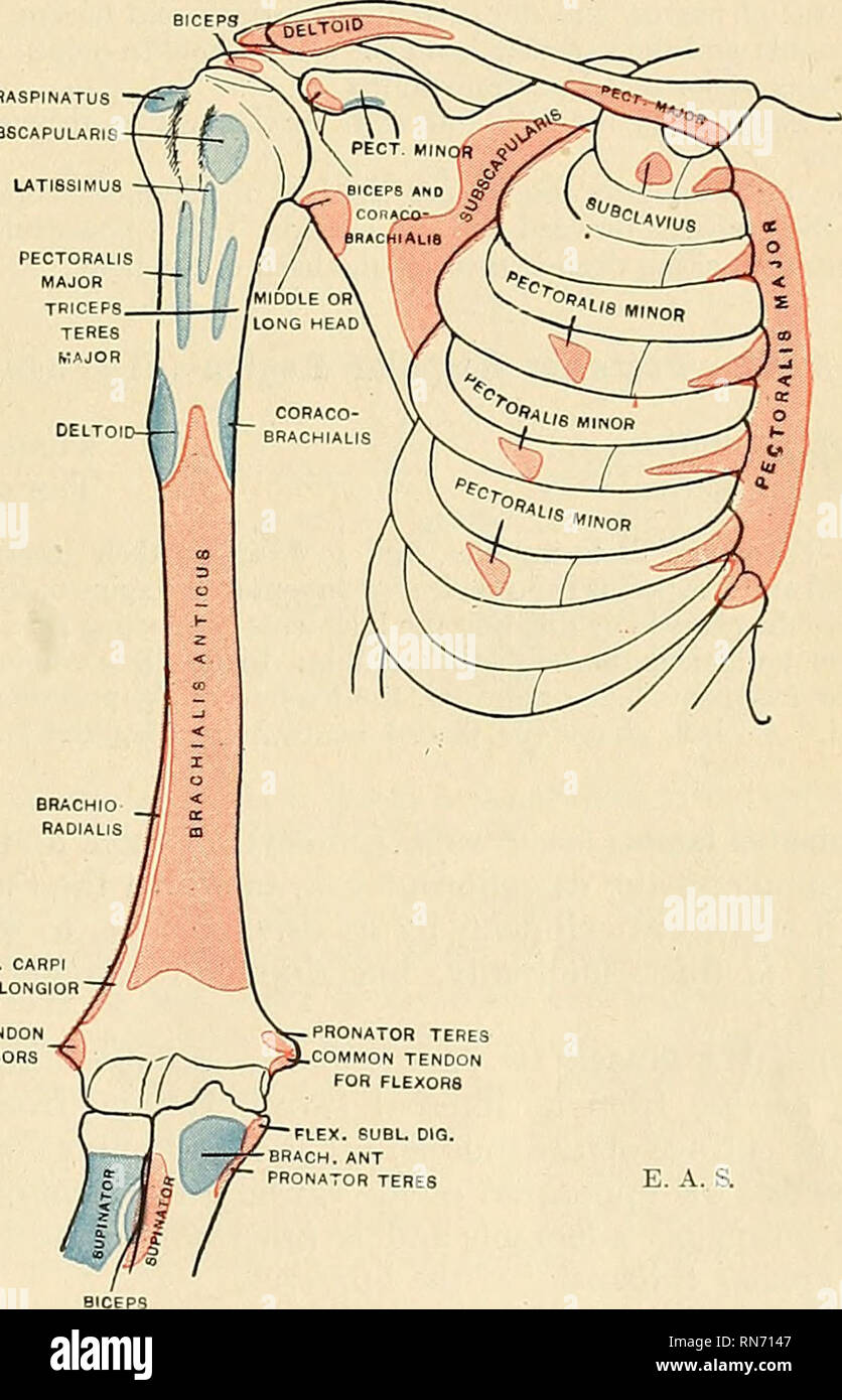
 Muscles Of The Pectoral Girdle And Upper Limbs Anatomy And
May present with multi directional instability.
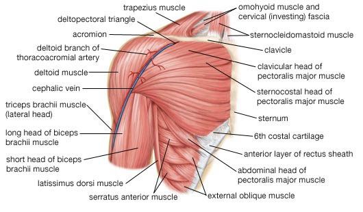
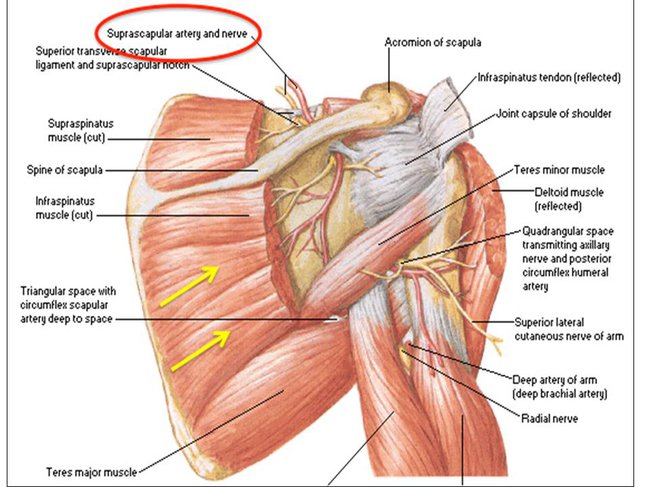
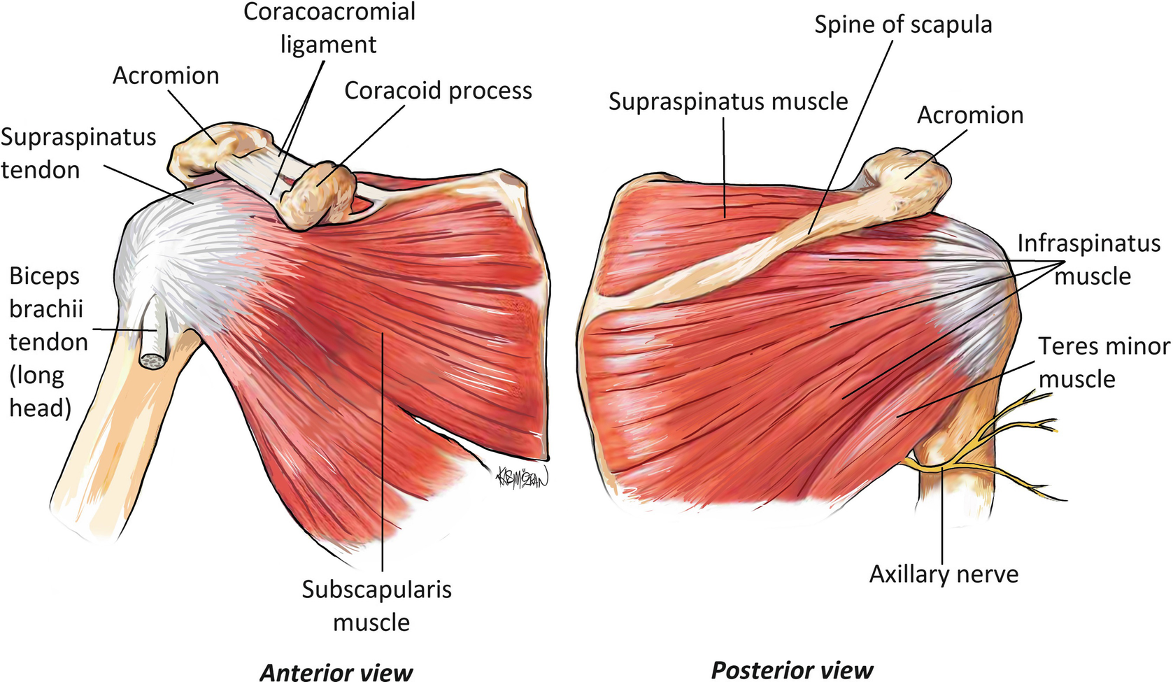
Posterior shoulder anatomy. The posterior shoulder with relatively fewer easily accessible arteries and branches is less frequently used. Boney structures shown are the acromion of the scapula the clavicle and the humerus. On the anterior side of the shoulder the coracobrachialis serratus anterior pectoralis major and pectoralis minor muscles work as a group to flex and adduct the scapula and humerus anteriorly toward the sternum.
There is a dissection assistance pdf file that you can use to assist you in your lab preparation. May have history of dislocation more likely recurrent posterior subluxation rps may be seen with seizures electrocution. The commonly used branches anteriorly include the thoracoacromial artery the lateral thoracic and the subscapular artery.
The shoulder joint also known as the glenohumeral joint is the main joint of the shoulder. Choose from 500 different sets of posterior shoulder anatomy flashcards on quizlet. Pain with adduction and internal rotation.
The shoulder joint ligaments shown are the acromioclavicular ligament coracoclavicular ligament the superior transverse scapular ligament and the joint capsule or glenohumeral ligaments. The acromion is a bony. Learn posterior shoulder anatomy with free interactive flashcards.
The muscles of the shoulder are associated with movements at the shoulder joint. The shoulder complex is composed of many different tissue types and it is the connective tissue that provides the supportive framework for the shoulders many functions. Uni directional instability posterior.
They produce the characteristic shape of the shoulder and can be divided into two groups. The different types of connective tissues in the shoulder are bone articular cartilage ligaments joint capsules and bursa see gross anatomy. The arterial anatomy of the shoulder is derived entirely from the axillary artery.
The latissimus dorsi and teres major on the posterior side extend and adduct the arm towards the vertebrae of the back. The shoulder is the group of structures in the region of the joint. Posterior shoulder the following video will walk you through the six steps to dissecting the posterior shoulder.
Other important bones in the shoulder include. The shoulder joint is formed where the humerus upper arm bone fits into the scapula shoulder blade like a ball and socket.
 Ultrasound Leadership Academy Intro To Shoulder Evaluation
 10 18 Anatomy Posterior Shoulder Musculoskeletal Block
 Technique Tuesday Posterior Shoulder Strength Peak
 Stabilizing The Shoulder Blade Joint Squat University
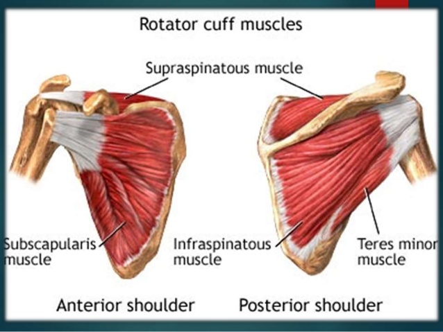
 Rotator Cuff Muscles Anterior And Posterior Shoulder
 Anatomy Of The Rtc Tendons Right Shoulder Download
 Boost Your Shoulder Health For Bigger Stronger Delts
 Judet Approach To Scapula Approaches Orthobullets
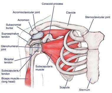 Shoulder Joint Anatomy Overview Gross Anatomy Microscopic
 Anatomy Musculoskeletal Ultrasonography
 Ucsd S Practical Guide To Clinical Medicine
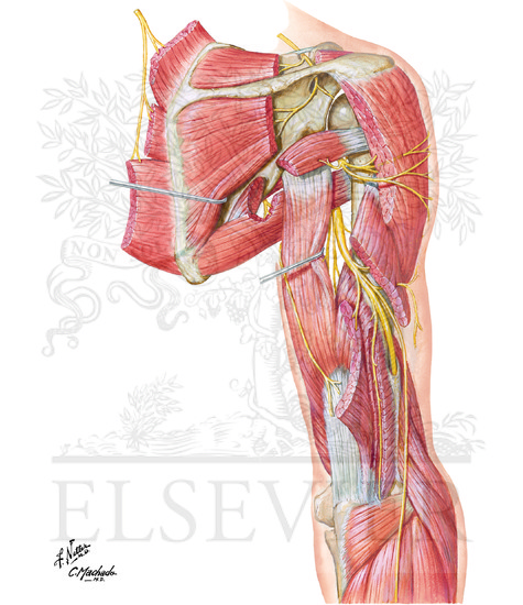 Radial Nerve In Arm And Nerves Of Posterior Shoulder
 Anterior And Posterior Anatomy Of The Right Shoulder
 Surface Anatomy Advanced Anatomy 2nd Ed
 Anterior And Posterior Aspects Of The Chest Back And
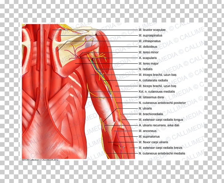 Serratus Anterior Muscle Serratus Posterior Inferior Muscle
 Rotator Cuff Anatomy Posterior Download Scientific Diagram
 Long Thoracic Nerve Injury The Shortest Route To Recovery
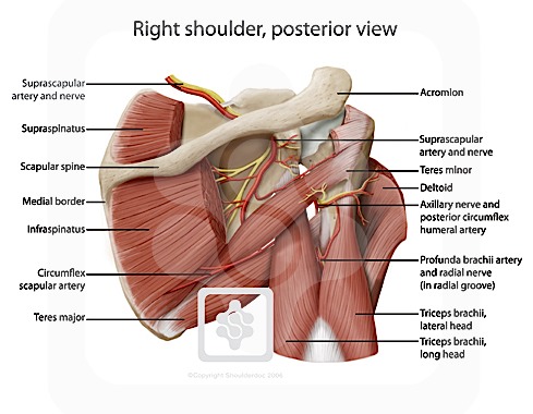 Nerves Of The Shoulder Shoulderdoc By Prof Lennard Funk
 Plyopic Massage Balls Set Neck Shoulders And Upper Back
 Anatomy Descriptive And Applied Anatomy The Anterior
Shoulder Dislocation The Cunningham Technique Department



Belum ada Komentar untuk "Posterior Shoulder Anatomy"
Posting Komentar